 Movie
Movie Controller


+ Open data

- Basic information
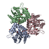
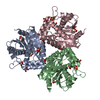
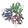
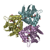
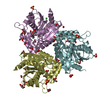
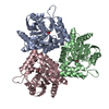
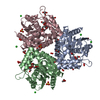
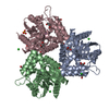
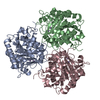
| Entry | Database: PDB / ID: 1a88 | ||||||
|---|---|---|---|---|---|---|---|
| Title | CHLOROPEROXIDASE L | ||||||
 Components Components | CHLOROPEROXIDASE L | ||||||
 Keywords Keywords | HALOPEROXIDASE / OXIDOREDUCTASE | ||||||
| Function / homology |  Function and homology information Function and homology informationchloride peroxidase activity / Oxidoreductases; Acting on a peroxide as acceptor; Peroxidases Similarity search - Function | ||||||
| Biological species |  Streptomyces lividans (bacteria) Streptomyces lividans (bacteria) | ||||||
| Method |  X-RAY DIFFRACTION / SYNCHROTRON / MOLECULAR REPLACEMENT / Resolution: 1.9 Å X-RAY DIFFRACTION / SYNCHROTRON / MOLECULAR REPLACEMENT / Resolution: 1.9 Å | ||||||
 Authors Authors | Hofmann, B. / Toelzer, S. / Pelletier, I. / Altenbuchner, J. / Van Pee, K.-H. / Hecht, H.-J. | ||||||
 Citation Citation | Journal: J.Mol.Biol. / Year: 1998 Title: Structural investigation of the cofactor-free chloroperoxidases. Authors: Hofmann, B. / Tolzer, S. / Pelletier, I. / Altenbuchner, J. / van Pee, K.H. / Hecht, H.J. | ||||||
| History |
|
- Structure visualization
| Structure viewer | Molecule: MolmilJmol/JSmol |
|---|
- Downloads & links
-Download
| PDBx/mmCIF format | 1a88.cif.gz | 174 KB | Display | PDBx/mmCIF format |
|---|---|---|---|---|
| PDB format | pdb1a88.ent.gz | 140 KB | Display | PDB format |
| PDBx/mmJSON format | 1a88.json.gz | Tree view | PDBx/mmJSON format | |
| Others |  Other downloads Other downloads |
-Validation report
| Arichive directory | https://data.pdbj.org/pub/pdb/validation_reports/a8/1a88ftp://data.pdbj.org/pub/pdb/validation_reports/a8/1a88 | HTTPS FTP |
|---|
-Related structure data
| Related structure data | 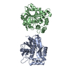 1a7uC 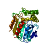 1a8qSC 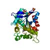 1a8sC 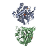 1a8uC 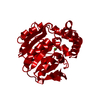 1brtC S: Starting model for refinement C: citing same article ( |
|---|---|
| Similar structure data |

-Links
 PDBj
PDBj


- Assembly
| Deposited unit | 
| ||||||||||||
|---|---|---|---|---|---|---|---|---|---|---|---|---|---|
| 1 |
| ||||||||||||
| Unit cell |
| ||||||||||||
| Noncrystallographic symmetry (NCS) | NCS oper:
|
-Components
| #1: Protein | Mass: 29816.207 Da / Num. of mol.: 3 Source method: isolated from a genetically manipulated source Source: (gene. exp.) Streptomyces lividans (bacteria) / Strain: TK64 / Gene: CPO-L / Plasmid: PRB882 / Gene (production host): CPO-L / Production host: Streptomyces aureofaciens (bacteria) / Strain (production host): TUE24-88 / References: UniProt: P49323, chloride peroxidase#2: Water | ChemComp-HOH / |  Mass: 18.015 Da / Num. of mol.: 507 / Source method: isolated from a natural source / Formula: H2O Mass: 18.015 Da / Num. of mol.: 507 / Source method: isolated from a natural source / Formula: H2O |
|---|
-Experimental details
-Experiment
| Experiment | Method: X-RAY DIFFRACTION / Number of used crystals: 1 |
|---|
- Sample preparation
| Crystal | Density Matthews: 2.79 Å3/Da / Density % sol: 55.3 % | |||||||||||||||
|---|---|---|---|---|---|---|---|---|---|---|---|---|---|---|---|---|
| Crystal grow | pH: 8.2 / Details: 2.0 M AMMONIUM SULFATE 100MM TRIS/HCL PH 8.2 | |||||||||||||||
| Crystal grow | *PLUS Temperature: 25 ℃ / Method: vapor diffusion, sitting drop | |||||||||||||||
| Components of the solutions | *PLUS
|
-Data collection
| Diffraction | Mean temperature: 283 K |
|---|---|
| Diffraction source | Source: SYNCHROTRON / Site: MPG/DESY, HAMBURG  / Beamline: BW6 / Wavelength: 1.1 / Beamline: BW6 / Wavelength: 1.1 |
| Detector | Type: MARRESEARCH / Detector: IMAGE PLATE / Date: Mar 1, 1995 / Details: MIRROR |
| Radiation | Monochromator: SI(111) / Monochromatic (M) / Laue (L): M / Scattering type: x-ray |
| Radiation wavelength | Wavelength: 1.1 Å / Relative weight: 1 |
| Reflection | Resolution: 1.9→44.2 Å / Num. obs: 65934 / % possible obs: 84.7 % / Observed criterion σ(I): 0 / Redundancy: 2 % / Biso Wilson estimate: 16.2 Å2 / Rmerge(I) obs: 0.078 / Net I/σ(I): 13.4 |
| Reflection shell | Resolution: 1.9→1.99 Å / Redundancy: 1.5 % / Rmerge(I) obs: 0.205 / Mean I/σ(I) obs: 5.2 / % possible all: 74.4 |
| Reflection | *PLUS Lowest resolution: 130 Å |
| Reflection shell | *PLUS % possible obs: 74.4 % |
- Processing
| Software |
| ||||||||||||||||||||||||||||||||||||||||||||||||||||||||||||||||||||||||||||||||||||
|---|---|---|---|---|---|---|---|---|---|---|---|---|---|---|---|---|---|---|---|---|---|---|---|---|---|---|---|---|---|---|---|---|---|---|---|---|---|---|---|---|---|---|---|---|---|---|---|---|---|---|---|---|---|---|---|---|---|---|---|---|---|---|---|---|---|---|---|---|---|---|---|---|---|---|---|---|---|---|---|---|---|---|---|---|---|
| Refinement | Method to determine structure: MOLECULAR REPLACEMENT Starting model: PDB ENTRY 1A8Q Resolution: 1.9→130 Å / Cross valid method: THROUGHOUT / σ(F): 0
| ||||||||||||||||||||||||||||||||||||||||||||||||||||||||||||||||||||||||||||||||||||
| Displacement parameters | Biso mean: 22.4 Å2 | ||||||||||||||||||||||||||||||||||||||||||||||||||||||||||||||||||||||||||||||||||||
| Refinement step | Cycle: LAST / Resolution: 1.9→130 Å
| ||||||||||||||||||||||||||||||||||||||||||||||||||||||||||||||||||||||||||||||||||||
| Refine LS restraints |
| ||||||||||||||||||||||||||||||||||||||||||||||||||||||||||||||||||||||||||||||||||||
| Software | *PLUS Name: REFMAC / Classification: refinement | ||||||||||||||||||||||||||||||||||||||||||||||||||||||||||||||||||||||||||||||||||||
| Refinement | *PLUS % reflection Rfree: 5 % / Rfactor obs: 0.16 / Rfactor Rwork: 0.16 | ||||||||||||||||||||||||||||||||||||||||||||||||||||||||||||||||||||||||||||||||||||
| Solvent computation | *PLUS | ||||||||||||||||||||||||||||||||||||||||||||||||||||||||||||||||||||||||||||||||||||
| Displacement parameters | *PLUS |
