 Movie
Movie Controller

[English] 日本語
 Yorodumi
- PDB-3hi4: Switching catalysis from hydrolysis to perhydrolysis in P. fluore... -
+ Open data

- Basic information
| Entry | Database: PDB / ID: 3hi4 | ||||||
|---|---|---|---|---|---|---|---|
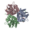
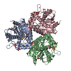
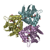
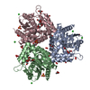
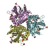
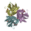
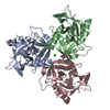
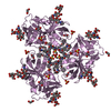
| Title | Switching catalysis from hydrolysis to perhydrolysis in P. fluorescens esterase | ||||||
 Components Components | Arylesterase | ||||||
 Keywords Keywords | HYDROLASE / esterase / hydrolysis / perhydrolysis / Peroxidase | ||||||
| Function / homology |  Function and homology information Function and homology informationarylesterase / Oxidoreductases / arylesterase activity / peroxidase activity Similarity search - Function | ||||||
| Biological species |  Pseudomonas fluorescens (bacteria) Pseudomonas fluorescens (bacteria) | ||||||
| Method |  X-RAY DIFFRACTION / MOLECULAR REPLACEMENT / Resolution: 2.25 Å X-RAY DIFFRACTION / MOLECULAR REPLACEMENT / Resolution: 2.25 Å | ||||||
 Authors Authors | Purpero, V.M. | ||||||
 Citation Citation | Journal: Biochemistry / Year: 2010 Title: Switching catalysis from hydrolysis to perhydrolysis in Pseudomonas fluorescens esterase. Authors: Yin, D.L.T. / Bernhardt, P. / Morley, K.L. / Jiang, Y. / Cheeseman, J.D. / Purpero, V. / Schrag, J.D. / Kazlauskas, R.J. | ||||||
| History |
|
- Structure visualization
| Structure viewer | Molecule: MolmilJmol/JSmol |
|---|
- Downloads & links
-Download
| PDBx/mmCIF format | 3hi4.cif.gz | 345.6 KB | Display | PDBx/mmCIF format |
|---|---|---|---|---|
| PDB format | pdb3hi4.ent.gz | 280.9 KB | Display | PDB format |
| PDBx/mmJSON format | 3hi4.json.gz | Tree view | PDBx/mmJSON format | |
| Others |  Other downloads Other downloads |
-Validation report
| Arichive directory | https://data.pdbj.org/pub/pdb/validation_reports/hi/3hi4ftp://data.pdbj.org/pub/pdb/validation_reports/hi/3hi4 | HTTPS FTP |
|---|
-Related structure data
| Related structure data |  3heaC 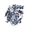 1va4S S: Starting model for refinement C: citing same article ( |
|---|---|
| Similar structure data |


-Links
 PDBj
PDBj


- Assembly
| Deposited unit | 
| ||||||||
|---|---|---|---|---|---|---|---|---|---|
| 1 | 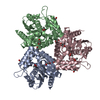
| ||||||||
| 2 | 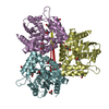
| ||||||||
| Unit cell |
|
-Components
| #1: Protein | Mass: 29977.912 Da / Num. of mol.: 6 / Mutation: L29P Source method: isolated from a genetically manipulated source Source: (gene. exp.) Pseudomonas fluorescens (bacteria) / Plasmid: pJOE2792 / Production host: #2: Chemical | ChemComp-ACT /   Mass: 59.044 Da / Num. of mol.: 10 / Source method: obtained synthetically / Formula: C2H3O2 Mass: 59.044 Da / Num. of mol.: 10 / Source method: obtained synthetically / Formula: C2H3O2#3: Chemical | ChemComp-SO4 /   Mass: 96.063 Da / Num. of mol.: 5 / Source method: obtained synthetically / Formula: SO4 Mass: 96.063 Da / Num. of mol.: 5 / Source method: obtained synthetically / Formula: SO4#4: Chemical | ChemComp-GOL /   Mass: 92.094 Da / Num. of mol.: 20 / Source method: obtained synthetically / Formula: C3H8O3 Mass: 92.094 Da / Num. of mol.: 20 / Source method: obtained synthetically / Formula: C3H8O3#5: Water | ChemComp-HOH / |  Mass: 18.015 Da / Num. of mol.: 1036 / Source method: isolated from a natural source / Formula: H2O Mass: 18.015 Da / Num. of mol.: 1036 / Source method: isolated from a natural source / Formula: H2O |
|---|
-Experimental details
-Experiment
| Experiment | Method: X-RAY DIFFRACTION / Number of used crystals: 1 |
|---|
- Sample preparation
| Crystal | Density Matthews: 4.42 Å3/Da / Density % sol: 72.15 % |
|---|---|
| Crystal grow | Temperature: 291 K / Method: vapor diffusion, hanging drop / pH: 7 Details: 1.75M ammonium sulfate 1% PEG 400, 0.1M HEPES pH 7.0, VAPOR DIFFUSION, HANGING DROP, temperature 291K |
-Data collection
| Diffraction | Mean temperature: 120 K |
|---|---|
| Diffraction source | Source: ROTATING ANODE / Type: RIGAKU MICROMAX-007 HF / Wavelength: 1.54 Å |
| Detector | Type: RIGAKU RAXIS IV++ / Detector: IMAGE PLATE / Date: Dec 8, 2008 / Details: mirrors |
| Radiation | Protocol: SINGLE WAVELENGTH / Monochromatic (M) / Laue (L): M / Scattering type: x-ray |
| Radiation wavelength | Wavelength: 1.54 Å / Relative weight: 1 |
| Reflection | Resolution: 2.25→39.97 Å / Num. obs: 146673 / % possible obs: 99.6 % / Observed criterion σ(I): 0 / Redundancy: 2.7 % / Biso Wilson estimate: 37.652 Å2 / Rsym value: 0.079 / Net I/σ(I): 12.3 |
| Reflection shell | Resolution: 2.25→2.29 Å / Redundancy: 2.5 % / Mean I/σ(I) obs: 2.15 / Num. unique all: 7291 / Rsym value: 0.356 / % possible all: 99.6 |
- Processing
| Software |
| ||||||||||||||||||||
|---|---|---|---|---|---|---|---|---|---|---|---|---|---|---|---|---|---|---|---|---|---|
| Refinement | Method to determine structure: MOLECULAR REPLACEMENT Starting model: 1VA4 Resolution: 2.25→39.97 Å / SU B: 3.939 / SU ML: 0.097 / Cross valid method: THROUGHOUT / ESU R: 0.155 / ESU R Free: 0.147 / Details: HYDROGENS HAVE BEEN ADDED IN THE RIDING POSITIONS
| ||||||||||||||||||||
| Displacement parameters | Biso mean: 29.392 Å2
| ||||||||||||||||||||
| Refinement step | Cycle: LAST / Resolution: 2.25→39.97 Å
| ||||||||||||||||||||
| Refine LS restraints |
| ||||||||||||||||||||
| LS refinement shell | Resolution: 2.25→2.29 Å
|
