 Movie
Movie Controller

[English] 日本語
 Yorodumi
- PDB-1zoi: Crystal Structure of a Stereoselective Esterase from Pseudomonas ... -
+ Open data

- Basic information
| Entry | Database: PDB / ID: 1zoi | ||||||
|---|---|---|---|---|---|---|---|
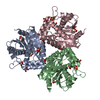
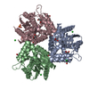
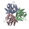
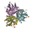
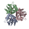
| Title | Crystal Structure of a Stereoselective Esterase from Pseudomonas putida IFO12996 | ||||||
 Components Components | esterase | ||||||
 Keywords Keywords | HYDROLASE / Esterase / Pseudomonas putida / alpha/beta hydrolase fold | ||||||
| Function / homology | : / alpha/beta hydrolase fold / Alpha/beta hydrolase fold-1 / Alpha/Beta hydrolase fold, catalytic domain / Alpha/Beta hydrolase fold / Rossmann fold / 3-Layer(aba) Sandwich / Alpha Beta / Esterase Function and homology information Function and homology information | ||||||
| Biological species |  Pseudomonas putida (bacteria) Pseudomonas putida (bacteria) | ||||||
| Method |  X-RAY DIFFRACTION / SYNCHROTRON / MOLECULAR REPLACEMENT / Resolution: 1.6 Å X-RAY DIFFRACTION / SYNCHROTRON / MOLECULAR REPLACEMENT / Resolution: 1.6 Å | ||||||
 Authors Authors | Elmi, F. / Lee, H.T. / Huang, J.Y. / Hsieh, Y.C. / Wang, Y.L. / Chen, Y.J. / Shaw, S.Y. / Chen, C.J. | ||||||
 Citation Citation | Journal: J.Bacteriol. / Year: 2005 Title: Stereoselective esterase from Pseudomonas putida IFO12996 reveals alpha/beta hydrolase folds for D-beta-acetylthioisobutyric acid synthesis Authors: Elmi, F. / Lee, H.T. / Huang, J.Y. / Hsieh, Y.C. / Wang, Y.L. / Chen, Y.J. / Shaw, S.Y. / Chen, C.J. | ||||||
| History |
|
- Structure visualization
| Structure viewer | Molecule: MolmilJmol/JSmol |
|---|
- Downloads & links
-Download
| PDBx/mmCIF format | 1zoi.cif.gz | 167.9 KB | Display | PDBx/mmCIF format |
|---|---|---|---|---|
| PDB format | pdb1zoi.ent.gz | 135.8 KB | Display | PDB format |
| PDBx/mmJSON format | 1zoi.json.gz | Tree view | PDBx/mmJSON format | |
| Others |  Other downloads Other downloads |
-Validation report
| Arichive directory | https://data.pdbj.org/pub/pdb/validation_reports/zo/1zoiftp://data.pdbj.org/pub/pdb/validation_reports/zo/1zoi | HTTPS FTP |
|---|
-Related structure data
| Similar structure data |
|---|


-Links
 PDBj
PDBj


- Assembly
| Deposited unit | 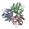
| ||||||||
|---|---|---|---|---|---|---|---|---|---|
| 1 |
| ||||||||
| Unit cell |
| ||||||||
| Details | The biological assembly is a homotrimer. |
-Components
| #1: Protein | Mass: 30201.867 Da / Num. of mol.: 3 Source method: isolated from a genetically manipulated source Source: (gene. exp.) Pseudomonas putida (bacteria) / Production host: References: UniProt: Q3HWU8, Hydrolases; Acting on ester bonds #2: Water | ChemComp-HOH / |  Mass: 18.015 Da / Num. of mol.: 317 / Source method: isolated from a natural source / Formula: H2O Mass: 18.015 Da / Num. of mol.: 317 / Source method: isolated from a natural source / Formula: H2O |
|---|
-Experimental details
-Experiment
| Experiment | Method: X-RAY DIFFRACTION / Number of used crystals: 1 |
|---|
- Sample preparation
| Crystal | Density Matthews: 2.091 Å3/Da / Density % sol: 40.09 % |
|---|---|
| Crystal grow | Temperature: 291 K / Method: vapor diffusion, hanging drop / pH: 4 Details: ammonium sulphate, citric acid, pH 4.0, VAPOR DIFFUSION, HANGING DROP, temperature 291K |
-Data collection
| Diffraction | Mean temperature: 100 K |
|---|---|
| Diffraction source | Source: SYNCHROTRON / Site: SPring-8  / Beamline: BL12B2 / Wavelength: 1 Å / Beamline: BL12B2 / Wavelength: 1 Å |
| Detector | Type: ADSC QUANTUM 4 / Detector: CCD / Date: Oct 27, 2004 |
| Radiation | Monochromator: Si 111 CHANNEL / Protocol: SINGLE WAVELENGTH / Monochromatic (M) / Laue (L): M / Scattering type: x-ray |
| Radiation wavelength | Wavelength: 1 Å / Relative weight: 1 |
| Reflection | Resolution: 1.6→25 Å / Num. all: 97023 / Num. obs: 93768 / % possible obs: 92.3 % / Observed criterion σ(F): 2 / Observed criterion σ(I): 2 / Redundancy: 6.3 % |
| Reflection shell | Resolution: 1.6→1.66 Å / % possible all: 82.3 |
- Processing
| Software |
| |||||||||||||||||||||
|---|---|---|---|---|---|---|---|---|---|---|---|---|---|---|---|---|---|---|---|---|---|---|
| Refinement | Method to determine structure: MOLECULAR REPLACEMENT / Resolution: 1.6→25 Å / σ(F): 2 / Stereochemistry target values: Engh & Huber
| |||||||||||||||||||||
| Refine analyze |
| |||||||||||||||||||||
| Refinement step | Cycle: LAST / Resolution: 1.6→25 Å
| |||||||||||||||||||||
| Refine LS restraints |
|
