 ムービー
ムービー コントローラー


+ データを開く

- 基本情報
基本情報
| 登録情報 | データベース: PDB / ID: 6nq1 | |||||||||||||||
|---|---|---|---|---|---|---|---|---|---|---|---|---|---|---|---|---|
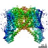
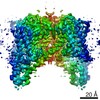
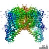
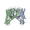
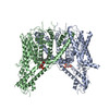
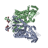
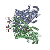
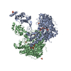
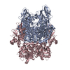
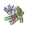
| タイトル | Cryo-EM structure of human TPC2 channel in the apo state | |||||||||||||||
 要素 要素 | Two pore calcium channel protein 2 | |||||||||||||||
 キーワード キーワード |  TRANSPORT PROTEIN (運搬体タンパク質) / channel / lysosome (リソソーム) TRANSPORT PROTEIN (運搬体タンパク質) / channel / lysosome (リソソーム) | |||||||||||||||
| 機能・相同性 |  機能・相同性情報 機能・相同性情報endosome to lysosome transport of low-density lipoprotein particle / negative regulation of developmental pigmentation / intracellular pH reduction / intracellularly phosphatidylinositol-3,5-bisphosphate-gated monatomic cation channel activity / ligand-gated sodium channel activity / NAADP-sensitive calcium-release channel activity / melanosome membrane / regulation of exocytosis / response to vitamin D / endolysosome membrane ...endosome to lysosome transport of low-density lipoprotein particle / negative regulation of developmental pigmentation / intracellular pH reduction / intracellularly phosphatidylinositol-3,5-bisphosphate-gated monatomic cation channel activity / ligand-gated sodium channel activity / NAADP-sensitive calcium-release channel activity / melanosome membrane / regulation of exocytosis / response to vitamin D / endolysosome membrane / phosphatidylinositol-3,5-bisphosphate binding / monoatomic ion channel complex / lysosome organization / sodium ion transmembrane transport / smooth muscle contraction / voltage-gated calcium channel activity / release of sequestered calcium ion into cytosol / monoatomic ion transmembrane transport / regulation of autophagy / calcium-mediated signaling / calcium channel activity / endocytosis involved in viral entry into host cell / intracellular calcium ion homeostasis / Stimuli-sensing channels / late endosome membrane / receptor-mediated endocytosis of virus by host cell / リソソーム / endosome membrane / lysosomal membrane / protein kinase binding / identical protein binding / 細胞質基質類似検索 - 分子機能 | |||||||||||||||
| 生物種 |  Homo sapiens (ヒト) Homo sapiens (ヒト) | |||||||||||||||
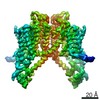
| 手法 | 電子顕微鏡法 / 単粒子再構成法 / クライオ電子顕微鏡法 / 解像度: 3.5 Å | |||||||||||||||
 データ登録者 データ登録者 | She, J. / Zeng, W. / Guo, J. / Chen, Q. / Bai, X. / Jiang, Y. | |||||||||||||||
| 資金援助 |  米国, 4件 米国, 4件
| |||||||||||||||
 引用 引用 | ジャーナル: Elife / 年: 2019 タイトル: Structural mechanisms of phospholipid activation of the human TPC2 channel. 著者: Ji She / Weizhong Zeng / Jiangtao Guo / Qingfeng Chen / Xiao-Chen Bai / Youxing Jiang /  要旨: Mammalian two-pore channels (TPCs) regulate the physiological functions of the endolysosome. Here we present cryo-EM structures of human TPC2 (HsTPC2), a phosphatidylinositol 3,5-bisphosphate (PI(3,5) ...Mammalian two-pore channels (TPCs) regulate the physiological functions of the endolysosome. Here we present cryo-EM structures of human TPC2 (HsTPC2), a phosphatidylinositol 3,5-bisphosphate (PI(3,5)P)-activated, Na selective channel, in the ligand-bound and apo states. The apo structure captures the closed conformation, while the ligand-bound form features the channel in both open and closed conformations. Combined with functional analysis, these structures provide insights into the mechanism of PI(3,5)P-regulated gating of TPC2, which is distinct from that of TPC1. Specifically, the endolysosome-specific PI(3,5)P binds at the first 6-TM and activates the channel - independently of the membrane potential - by inducing a structural change at the pore-lining inner helix (IS6), which forms a continuous helix in the open state but breaks into two segments at Gly317 in the closed state. Additionally, structural comparison to the voltage-dependent TPC1 structure allowed us to identify Ile551 as being responsible for the loss of voltage dependence in TPC2. | |||||||||||||||
| 履歴 |
|
- 構造の表示
構造の表示
| ムービー |
ムービービューア |
|---|---|
| 構造ビューア | 分子: MolmilJmol/JSmol |
- ダウンロードとリンク
-ダウンロード
| PDBx/mmCIF形式 | 6nq1.cif.gz | 222.8 KB | 表示 | PDBx/mmCIF形式 |
|---|---|---|---|---|
| PDB形式 | pdb6nq1.ent.gz | 184.5 KB | 表示 | PDB形式 |
| PDBx/mmJSON形式 | 6nq1.json.gz | ツリー表示 | PDBx/mmJSON形式 | |
| その他 |  その他のダウンロード その他のダウンロード |
-検証レポート
| アーカイブディレクトリ | https://data.pdbj.org/pub/pdb/validation_reports/nq/6nq1ftp://data.pdbj.org/pub/pdb/validation_reports/nq/6nq1 | HTTPS FTP |
|---|
-関連構造データ


-リンク
 PDBj
PDBj
- 集合体
集合体
| 登録構造単位 | 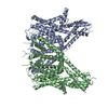
|
|---|---|
| 1 |
|
-要素
| #1: タンパク質 | 分子量: 85671.828 Da / 分子数: 2 / 由来タイプ: 組換発現 / 由来: (組換発現) Homo sapiens (ヒト) / 遺伝子: TPCN2, TPC2 / 発現宿主: Homo sapiens (ヒト) / 参照: UniProt: Q8NHX9 |
|---|
-実験情報
-実験
| 実験 | 手法: 電子顕微鏡法 |
|---|---|
| EM実験 | 試料の集合状態: PARTICLE / 3次元再構成法: 単粒子再構成法 |
- 試料調製
試料調製
| 構成要素 | 名称: Two-pore channel 2 / タイプ: COMPLEX / Entity ID: all / 由来: RECOMBINANT |
|---|---|
| 由来(天然) | 生物種: Homo sapiens (ヒト) |
| 由来(組換発現) | 生物種: Homo sapiens (ヒト) |
| 緩衝液 | pH: 8 |
| 試料 | 包埋: NO / シャドウイング: NO / 染色: NO / 凍結: YES |
| 試料支持 | 詳細: unspecified |
| 急速凍結 | 凍結剤: ETHANE |
- 電子顕微鏡撮影
| 実験機器 |  モデル: Titan Krios / 画像提供: FEI Company |
|---|---|
| 顕微鏡 | モデル: FEI TITAN KRIOS |
| 電子銃 | 電子線源: FIELD EMISSION GUN / 加速電圧: 300 kV / 照射モード: FLOOD BEAM |
| 電子レンズ | モード: BRIGHT FIELDBright-field microscopy |
| 撮影 | 電子線照射量: 1.6 e/Å2 フィルム・検出器のモデル: GATAN K2 SUMMIT (4k x 4k) |
- 解析
解析
| ソフトウェア | 名称: PHENIX / バージョン: 1.14_3260: / 分類: 精密化 |
|---|---|
| CTF補正 | タイプ: NONE |
| 3次元再構成 | 解像度: 3.5 Å / 解像度の算出法: FSC 0.143 CUT-OFF / 粒子像の数: 96361 / 対称性のタイプ: POINT |
