 Movie
Movie Controller

[English] 日本語
 Yorodumi
- PDB-6nq0: Cryo-EM structure of human TPC2 channel in the ligand-bound open state -
+ Open data

- Basic information
| Entry | Database: PDB / ID: 6nq0 | |||||||||||||||||||||||||||||||||
|---|---|---|---|---|---|---|---|---|---|---|---|---|---|---|---|---|---|---|---|---|---|---|---|---|---|---|---|---|---|---|---|---|---|---|
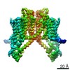
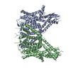
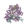
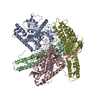
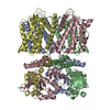
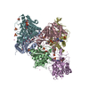
| Title | Cryo-EM structure of human TPC2 channel in the ligand-bound open state | |||||||||||||||||||||||||||||||||
 Components Components | Two pore calcium channel protein 2 | |||||||||||||||||||||||||||||||||
 Keywords Keywords | TRANSPORT PROTEIN / channel / lysosome | |||||||||||||||||||||||||||||||||
| Function / homology |  Function and homology information Function and homology informationendosome to lysosome transport of low-density lipoprotein particle / negative regulation of developmental pigmentation / intracellular pH reduction / intracellularly phosphatidylinositol-3,5-bisphosphate-gated monatomic cation channel activity / NAADP-sensitive calcium-release channel activity / regulation of exocytosis / melanosome membrane / endolysosome membrane / phosphatidylinositol-3,5-bisphosphate binding / response to vitamin D ...endosome to lysosome transport of low-density lipoprotein particle / negative regulation of developmental pigmentation / intracellular pH reduction / intracellularly phosphatidylinositol-3,5-bisphosphate-gated monatomic cation channel activity / NAADP-sensitive calcium-release channel activity / regulation of exocytosis / melanosome membrane / endolysosome membrane / phosphatidylinositol-3,5-bisphosphate binding / response to vitamin D / ligand-gated sodium channel activity / lysosome organization / monoatomic ion channel complex / smooth muscle contraction / voltage-gated calcium channel activity / receptor-mediated endocytosis of virus by host cell / release of sequestered calcium ion into cytosol / regulation of autophagy / sodium ion transmembrane transport / calcium-mediated signaling / Stimuli-sensing channels / calcium channel activity / intracellular calcium ion homeostasis / late endosome membrane / monoatomic ion transmembrane transport / lysosome / endosome membrane / endocytosis involved in viral entry into host cell / lysosomal membrane / protein kinase binding / identical protein binding / cytosol Similarity search - Function | |||||||||||||||||||||||||||||||||
| Biological species |  Homo sapiens (human) Homo sapiens (human) | |||||||||||||||||||||||||||||||||
| Method | ELECTRON MICROSCOPY / single particle reconstruction / cryo EM / Resolution: 3.7 Å | |||||||||||||||||||||||||||||||||
 Authors Authors | She, J. / Zeng, W. / Guo, J. / Chen, Q. / Bai, X. / Jiang, Y. | |||||||||||||||||||||||||||||||||
| Funding support |  United States, 4items United States, 4items
| |||||||||||||||||||||||||||||||||
 Citation Citation | Journal: Elife / Year: 2019 Title: Structural mechanisms of phospholipid activation of the human TPC2 channel. Authors: Ji She / Weizhong Zeng / Jiangtao Guo / Qingfeng Chen / Xiao-Chen Bai / Youxing Jiang /  Abstract: Mammalian two-pore channels (TPCs) regulate the physiological functions of the endolysosome. Here we present cryo-EM structures of human TPC2 (HsTPC2), a phosphatidylinositol 3,5-bisphosphate (PI(3,5) ...Mammalian two-pore channels (TPCs) regulate the physiological functions of the endolysosome. Here we present cryo-EM structures of human TPC2 (HsTPC2), a phosphatidylinositol 3,5-bisphosphate (PI(3,5)P)-activated, Na selective channel, in the ligand-bound and apo states. The apo structure captures the closed conformation, while the ligand-bound form features the channel in both open and closed conformations. Combined with functional analysis, these structures provide insights into the mechanism of PI(3,5)P-regulated gating of TPC2, which is distinct from that of TPC1. Specifically, the endolysosome-specific PI(3,5)P binds at the first 6-TM and activates the channel - independently of the membrane potential - by inducing a structural change at the pore-lining inner helix (IS6), which forms a continuous helix in the open state but breaks into two segments at Gly317 in the closed state. Additionally, structural comparison to the voltage-dependent TPC1 structure allowed us to identify Ile551 as being responsible for the loss of voltage dependence in TPC2. | |||||||||||||||||||||||||||||||||
| History |
|
- Structure visualization
| Movie |
Movie viewer |
|---|---|
| Structure viewer | Molecule: MolmilJmol/JSmol |
- Downloads & links
-Download
| PDBx/mmCIF format | 6nq0.cif.gz | 232.1 KB | Display | PDBx/mmCIF format |
|---|---|---|---|---|
| PDB format | pdb6nq0.ent.gz | 184.1 KB | Display | PDB format |
| PDBx/mmJSON format | 6nq0.json.gz | Tree view | PDBx/mmJSON format | |
| Others |  Other downloads Other downloads |
-Validation report
| Arichive directory | https://data.pdbj.org/pub/pdb/validation_reports/nq/6nq0ftp://data.pdbj.org/pub/pdb/validation_reports/nq/6nq0 | HTTPS FTP |
|---|
-Related structure data
| Related structure data | 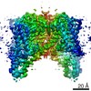 0477MC 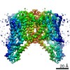 0478C 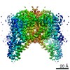 0479C 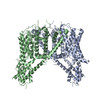 6nq1C 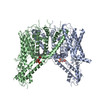 6nq2C M: map data used to model this data C: citing same article ( |
|---|---|
| Similar structure data |

-Links
 PDBj
PDBj
- Assembly
| Deposited unit | 
|
|---|---|
| 1 |
|
-Components
| #1: Protein | Mass: 85671.828 Da / Num. of mol.: 2 Source method: isolated from a genetically manipulated source Source: (gene. exp.) Homo sapiens (human) / Gene: TPCN2, TPC2 / Production host: Homo sapiens (human) / References: UniProt: Q8NHX9#2: Chemical |   Mass: 746.566 Da / Num. of mol.: 2 / Source method: obtained synthetically / Formula: C25H49O19P3 Mass: 746.566 Da / Num. of mol.: 2 / Source method: obtained synthetically / Formula: C25H49O19P3Has protein modification | Y | |
|---|
-Experimental details
-Experiment
| Experiment | Method: ELECTRON MICROSCOPY |
|---|---|
| EM experiment | Aggregation state: PARTICLE / 3D reconstruction method: single particle reconstruction |
- Sample preparation
| Component | Name: The complex of TPC2 with PI(3,5)P2 / Type: COMPLEX / Entity ID: #1 / Source: RECOMBINANT |
|---|---|
| Source (natural) | Organism: Homo sapiens (human) |
| Source (recombinant) | Organism: Homo sapiens (human) |
| Buffer solution | pH: 8 |
| Specimen | Embedding applied: NO / Shadowing applied: NO / Staining applied: NO / Vitrification applied: YES |
| Specimen support | Details: unspecified |
| Vitrification | Cryogen name: ETHANE |
- Electron microscopy imaging
| Experimental equipment |  Model: Titan Krios / Image courtesy: FEI Company |
|---|---|
| Microscopy | Model: FEI TITAN KRIOS |
| Electron gun | Electron source:  FIELD EMISSION GUN / Accelerating voltage: 300 kV / Illumination mode: FLOOD BEAM FIELD EMISSION GUN / Accelerating voltage: 300 kV / Illumination mode: FLOOD BEAM |
| Electron lens | Mode: BRIGHT FIELD |
| Image recording | Electron dose: 1.6 e/Å2 / Film or detector model: GATAN K2 SUMMIT (4k x 4k) |
- Processing
| Software | Name: PHENIX / Version: 1.14_3260: / Classification: refinement |
|---|---|
| EM software | Name: PHENIX / Category: model refinement |
| CTF correction | Type: NONE |
| 3D reconstruction | Resolution: 3.7 Å / Resolution method: FSC 0.143 CUT-OFF / Num. of particles: 33441 / Symmetry type: POINT |
