 Movie
Movie Controller

[English] 日本語
 Yorodumi
- PDB-6c9a: Cryo-EM structure of mouse TPC1 channel in the PtdIns(3,5)P2-boun... -
+ Open data

- Basic information
| Entry | Database: PDB / ID: 6c9a | ||||||||||||||||||||||||||||||||||||||||||||||||||||||
|---|---|---|---|---|---|---|---|---|---|---|---|---|---|---|---|---|---|---|---|---|---|---|---|---|---|---|---|---|---|---|---|---|---|---|---|---|---|---|---|---|---|---|---|---|---|---|---|---|---|---|---|---|---|---|---|
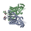
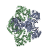
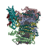
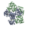
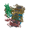
| Title | Cryo-EM structure of mouse TPC1 channel in the PtdIns(3,5)P2-bound state | ||||||||||||||||||||||||||||||||||||||||||||||||||||||
 Components Components | Two pore calcium channel protein 1 | ||||||||||||||||||||||||||||||||||||||||||||||||||||||
 Keywords Keywords | MEMBRANE PROTEIN / Ion channel | ||||||||||||||||||||||||||||||||||||||||||||||||||||||
| Function / homology |  Function and homology information Function and homology informationvoltage-gated channel activity / endolysosome / intracellularly phosphatidylinositol-3,5-bisphosphate-gated monatomic cation channel activity / NAADP-sensitive calcium-release channel activity / Stimuli-sensing channels / phosphatidylinositol-3,5-bisphosphate binding / syntaxin binding / voltage-gated sodium channel activity / ligand-gated sodium channel activity / monoatomic ion channel complex ...voltage-gated channel activity / endolysosome / intracellularly phosphatidylinositol-3,5-bisphosphate-gated monatomic cation channel activity / NAADP-sensitive calcium-release channel activity / Stimuli-sensing channels / phosphatidylinositol-3,5-bisphosphate binding / syntaxin binding / voltage-gated sodium channel activity / ligand-gated sodium channel activity / monoatomic ion channel complex / positive regulation of autophagy / sodium ion transmembrane transport / recycling endosome membrane / early endosome membrane / endosome membrane / endocytosis involved in viral entry into host cell / lysosomal membrane / protein homodimerization activity / membrane / identical protein binding Similarity search - Function | ||||||||||||||||||||||||||||||||||||||||||||||||||||||
| Biological species |  | ||||||||||||||||||||||||||||||||||||||||||||||||||||||
| Method | ELECTRON MICROSCOPY / single particle reconstruction / cryo EM / Resolution: 3.2 Å | ||||||||||||||||||||||||||||||||||||||||||||||||||||||
 Authors Authors | She, J. / Guo, J. / Chen, Q. / Bai, X. / Jiang, Y. | ||||||||||||||||||||||||||||||||||||||||||||||||||||||
| Funding support |  United States, 5items United States, 5items
| ||||||||||||||||||||||||||||||||||||||||||||||||||||||
 Citation Citation | Journal: Nature / Year: 2018 Title: Structural insights into the voltage and phospholipid activation of the mammalian TPC1 channel. Authors: Ji She / Jiangtao Guo / Qingfeng Chen / Weizhong Zeng / Youxing Jiang / Xiao-Chen Bai /  Abstract: The organellar two-pore channel (TPC) functions as a homodimer, in which each subunit contains two homologous Shaker-like six-transmembrane (6-TM)-domain repeats. TPCs belong to the voltage-gated ion ...The organellar two-pore channel (TPC) functions as a homodimer, in which each subunit contains two homologous Shaker-like six-transmembrane (6-TM)-domain repeats. TPCs belong to the voltage-gated ion channel superfamily and are ubiquitously expressed in animals and plants. Mammalian TPC1 and TPC2 are localized at the endolysosomal membrane, and have critical roles in regulating the physiological functions of these acidic organelles. Here we present electron cryo-microscopy structures of mouse TPC1 (MmTPC1)-a voltage-dependent, phosphatidylinositol 3,5-bisphosphate (PtdIns(3,5)P)-activated Na-selective channel-in both the apo closed state and ligand-bound open state. Combined with functional analysis, these structures provide comprehensive structural insights into the selectivity and gating mechanisms of mammalian TPC channels. The channel has a coin-slot-shaped ion pathway in the filter that defines the selectivity of mammalian TPCs. Only the voltage-sensing domain from the second 6-TM domain confers voltage dependence on MmTPC1. Endolysosome-specific PtdIns(3,5)P binds to the first 6-TM domain and activates the channel under conditions of depolarizing membrane potential. Structural comparisons between the apo and PtdIns(3,5)P-bound structures show the interplay between voltage and ligand in channel activation. These MmTPC1 structures reveal lipid binding and regulation in a 6-TM voltage-gated channel, which is of interest in light of the emerging recognition of the importance of phosphoinositide regulation of ion channels. | ||||||||||||||||||||||||||||||||||||||||||||||||||||||
| History |
|
- Structure visualization
| Movie |
Movie viewer |
|---|---|
| Structure viewer | Molecule: MolmilJmol/JSmol |
- Downloads & links
-Download
| PDBx/mmCIF format | 6c9a.cif.gz | 275.2 KB | Display | PDBx/mmCIF format |
|---|---|---|---|---|
| PDB format | pdb6c9a.ent.gz | 220.6 KB | Display | PDB format |
| PDBx/mmJSON format | 6c9a.json.gz | Tree view | PDBx/mmJSON format | |
| Others |  Other downloads Other downloads |
-Validation report
| Arichive directory | https://data.pdbj.org/pub/pdb/validation_reports/c9/6c9aftp://data.pdbj.org/pub/pdb/validation_reports/c9/6c9a | HTTPS FTP |
|---|
-Related structure data
| Related structure data |  7435MC  7434C  6c96C C: citing same article ( M: map data used to model this data |
|---|---|
| Similar structure data |


-Links
 PDBj
PDBj
- Assembly
| Deposited unit | 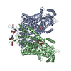
|
|---|---|
| 1 |
|
-Components
| #1: Protein | Mass: 95414.758 Da / Num. of mol.: 2 Source method: isolated from a genetically manipulated source Source: (gene. exp.)  Homo sapiens (human) / References: UniProt: Q9EQJ0 Homo sapiens (human) / References: UniProt: Q9EQJ0#2: Polysaccharide | Source method: isolated from a genetically manipulated source #3: Chemical |   Mass: 746.566 Da / Num. of mol.: 2 / Source method: obtained synthetically / Formula: C25H49O19P3 Mass: 746.566 Da / Num. of mol.: 2 / Source method: obtained synthetically / Formula: C25H49O19P3#4: Sugar |   Type: D-saccharide, beta linking / Mass: 221.208 Da / Num. of mol.: 2 Type: D-saccharide, beta linking / Mass: 221.208 Da / Num. of mol.: 2Source method: isolated from a genetically manipulated source Formula: C8H15NO6 Has protein modification | Y | |
|---|
-Experimental details
-Experiment
| Experiment | Method: ELECTRON MICROSCOPY |
|---|---|
| EM experiment | Aggregation state: PARTICLE / 3D reconstruction method: single particle reconstruction |
- Sample preparation
| Component | Name: PtdIns(3,5)P2-bound mouse TPC1 / Type: ORGANELLE OR CELLULAR COMPONENT / Entity ID: #1 / Source: RECOMBINANT |
|---|---|
| Source (natural) | Organism: |
| Source (recombinant) | Organism: Homo sapiens (human) / Cell: HEK293F / Plasmid: pEZT-BM |
| Buffer solution | pH: 8 |
| Specimen | Embedding applied: NO / Shadowing applied: NO / Staining applied: NO / Vitrification applied: YES |
| Vitrification | Cryogen name: ETHANE |
- Electron microscopy imaging
| Experimental equipment |  Model: Titan Krios / Image courtesy: FEI Company |
|---|---|
| Microscopy | Model: FEI TITAN KRIOS |
| Electron gun | Electron source:  FIELD EMISSION GUN / Accelerating voltage: 300 kV / Illumination mode: FLOOD BEAM FIELD EMISSION GUN / Accelerating voltage: 300 kV / Illumination mode: FLOOD BEAM |
| Electron lens | Mode: BRIGHT FIELD |
| Image recording | Electron dose: 50 e/Å2 / Film or detector model: GATAN K2 SUMMIT (4k x 4k) |
- Processing
| Software | Name: PHENIX / Version: 1.11.1_2575: / Classification: refinement | ||||||||||||||||||||||||
|---|---|---|---|---|---|---|---|---|---|---|---|---|---|---|---|---|---|---|---|---|---|---|---|---|---|
| EM software | Name: PHENIX / Category: model refinement | ||||||||||||||||||||||||
| CTF correction | Type: PHASE FLIPPING AND AMPLITUDE CORRECTION | ||||||||||||||||||||||||
| 3D reconstruction | Resolution: 3.2 Å / Resolution method: FSC 0.143 CUT-OFF / Num. of particles: 82819 / Symmetry type: POINT | ||||||||||||||||||||||||
| Refine LS restraints |
|
