[日本語] English
- PDB-2fxo: Structure of the human beta-myosin S2 fragment -

+
データを開く


IDまたはキーワード:

読み込み中...

-
基本情報

登録情報
データベース: PDB / ID: 2fxo
タイトルStructure of the human beta-myosin S2 fragment
要素Myosin heavy chain, cardiac muscle beta isoformミオシン
キーワードCONTRACTILE PROTEIN / COILED COIL (DIMERIC / PARALLEL) / FAMILIAL HYPERTROPHIC CARDIOMYOPATHY / FHC-ASSOCIATED MUTANT E924K / THICK FILAMENT (ミオフィラメント)
機能・相同性
機能・相同性情報


regulation of slow-twitch skeletal muscle fiber contraction / regulation of the force of skeletal muscle contraction / muscle myosin complex / muscle filament sliding / regulation of the force of heart contraction / transition between fast and slow fiber / myosin filament / myosin II complex / adult heart development / cardiac muscle hypertrophy in response to stress ...regulation of slow-twitch skeletal muscle fiber contraction / regulation of the force of skeletal muscle contraction / muscle myosin complex / muscle filament sliding / regulation of the force of heart contraction / transition between fast and slow fiber / myosin filament / myosin II complex / adult heart development / cardiac muscle hypertrophy in response to stress / myosin complex / sarcomere organization / microfilament motor activity / ventricular cardiac muscle tissue morphogenesis / myofibril / skeletal muscle contraction / striated muscle contraction / stress fiber / ATP metabolic process / cardiac muscle contraction / regulation of heart rate / sarcomere / muscle contraction / Z disc / actin filament binding / calmodulin binding / ATP binding / 細胞質
類似検索 - 分子機能
Single alpha-helices involved in coiled-coils or other helix-helix interfaces - #340 / DNA repair protein XRCC4-like, C-terminal / Myosin tail / Myosin tail / Myosin N-terminal SH3-like domain / Myosin S1 fragment, N-terminal / Myosin, N-terminal, SH3-like / Myosin N-terminal SH3-like domain profile. / Short calmodulin-binding motif containing conserved Ile and Gln residues. / Myosin head, motor domain ...Single alpha-helices involved in coiled-coils or other helix-helix interfaces - #340 / DNA repair protein XRCC4-like, C-terminal / Myosin tail / Myosin tail / Myosin N-terminal SH3-like domain / Myosin S1 fragment, N-terminal / Myosin, N-terminal, SH3-like / Myosin N-terminal SH3-like domain profile. / Short calmodulin-binding motif containing conserved Ile and Gln residues. / Myosin head, motor domain / Myosin head (motor domain) / Myosin motor domain profile. / Myosin. Large ATPases. / IQ motif profile. / IQ motif, EF-hand binding site / Kinesin motor domain superfamily / Single alpha-helices involved in coiled-coils or other helix-helix interfaces / Up-down Bundle / P-loop containing nucleoside triphosphate hydrolase / Mainly Alpha
類似検索 - ドメイン・相同性
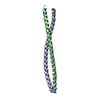
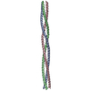
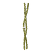
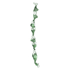
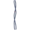
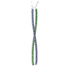
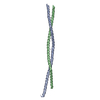
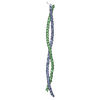
生物種Homo sapiens (ヒト)
手法X線回折 / シンクロトロン / SAD WITH SEMI-BRUTE- FORCE MOLECULAR REPLACEMENT / 解像度: 2.5 Å
データ登録者Blankenfeldt, W. / Thoma, N.H. / Wray, J.S. / Gautel, M. / Schlichting, I.
引用
ジャーナル: Proc Natl Acad Sci U S A / : 2006
タイトル: Crystal structures of human cardiac beta-myosin II S2-Delta provide insight into the functional role of the S2 subfragment.
著者: Wulf Blankenfeldt / Nicolas H Thomä / John S Wray / Mathias Gautel / Ilme Schlichting /
要旨: Myosin II is the major component of the muscle thick filament. It consists of two N-terminal S1 subfragments ("heads") connected to a long dimeric coiled-coil rod. The rod is in itself twofold ...Myosin II is the major component of the muscle thick filament. It consists of two N-terminal S1 subfragments ("heads") connected to a long dimeric coiled-coil rod. The rod is in itself twofold symmetric, but in the filament, the two heads point away from the filament surface and are therefore not equivalent. This breaking of symmetry requires the initial section of the rod, subfragment 2 (S2), to be relatively flexible. S2 is an important functional element, involved in various mechanisms by which the activity of smooth and striated muscle is regulated. We have determined crystal structures of the 126 N-terminal residues of S2 from human cardiac beta-myosin II (S2-Delta), of both WT and the disease-associated E924K mutant. S2-Delta is a straight parallel dimeric coiled coil, but the N terminus of one chain is disordered in WT-S2-Delta due to crystal contacts, indicative of unstable local structure. Bulky noncanonical side chains pack into a/d positions of S2-Delta's N terminus, leading to defined local asymmetry and axial stagger, which could induce nonequivalence of the S1 subfragments. Additionally, S2 possesses a conserved charge distribution with three prominent rings of negative potential within S2-Delta, the first of which may provide a binding interface for the "blocked head" of smooth muscle myosin in the OFF state. The observation that many disease-associated mutations affect the second negatively charged ring further suggests that charge interactions play an important role in regulation of cardiac muscle activity through myosin-binding protein C.
#1: ジャーナル: J.Mol.Biol. / : 1999
タイトル: Mutations in Beta-Myosin S2 that Cause Familial Hypertrophic Cardiomyopathy (Fhc) Abolish the Interaction with the Regulatory Domain of Myosin-Binding Protein-C
著者: Gruen, M. / Gautel, M.
#2: ジャーナル: Nature / : 2003
タイトル: Visualization of an Unstable Coiled Coil from the Scallop Myosin Rod
著者: Li, Y. / Brown, J.H. / Reshetnikova, L. / Blazsek, A. / Farkas, L. / Nyitray, L. / Cohen, C.
履歴
登録2006年2月6日登録サイト: RCSB / 処理サイト: PDBJ
改定 1.02006年11月21日Provider: repository / タイプ: Initial release
改定 1.12008年5月1日Group: Version format compliance
改定 1.22011年7月13日Group: Advisory / Version format compliance
改定 1.32017年10月18日Group: Advisory / Refinement description / カテゴリ: pdbx_unobs_or_zero_occ_atoms / software
Item: _software.classification / _software.name / _software.version
改定 1.42021年11月10日Group: Advisory / Database references
カテゴリ: database_2 / pdbx_unobs_or_zero_occ_atoms / struct_ref_seq_dif
Item: _database_2.pdbx_DOI / _database_2.pdbx_database_accession / _struct_ref_seq_dif.details
改定 1.52023年10月25日Group: Data collection / Refinement description
カテゴリ: chem_comp_atom / chem_comp_bond / pdbx_initial_refinement_model

-
構造の表示

構造ビューア分子:
MolmilJmol/JSmol

ダウンロードとリンク

-
集合体

登録構造単位
A: Myosin heavy chain, cardiac muscle beta isoform
B: Myosin heavy chain, cardiac muscle beta isoform
C: Myosin heavy chain, cardiac muscle beta isoform
D: Myosin heavy chain, cardiac muscle beta isoform


分子量 (理論値)分子数
合計 (水以外)60,4254
ポリマ-60,4254
非ポリマー00
0
1
A: Myosin heavy chain, cardiac muscle beta isoform
B: Myosin heavy chain, cardiac muscle beta isoform


分子量 (理論値)分子数
合計 (水以外)30,2122
ポリマ-30,2122
非ポリマー00
0
タイプ名称対称操作
identity operation1_555x,y,z1
Buried area6890 Å2
ΔGint-72 kcal/mol
Surface area18880 Å2
手法PISA
2
C: Myosin heavy chain, cardiac muscle beta isoform
D: Myosin heavy chain, cardiac muscle beta isoform


分子量 (理論値)分子数
合計 (水以外)30,2122
ポリマ-30,2122
非ポリマー00
0
タイプ名称対称操作
identity operation1_555x,y,z1
Buried area6840 Å2
ΔGint-72 kcal/mol
Surface area19050 Å2
手法PISA
単位格子
Length a, b, c (Å)40.157, 41.867, 97.795
Angle α, β, γ (deg.)91.11, 92.73, 107.18
Int Tables number1
Space group name H-MP1

-
要素

#1: タンパク質
Myosin heavy chain, cardiac muscle beta isoform / ミオシン / BETA-MYOSIN / MyHC-beta


分子量: 15106.145 Da / 分子数: 4 / 断片: DELTA-S2 FRAGMENT (838-963) / 変異: E924K / 由来タイプ: 組換発現 / 由来: (組換発現) Homo sapiens (ヒト) / 組織: HEART MUSCLE / 遺伝子: HSBMHC / プラスミド: PET / 生物種 (発現宿主): Escherichia coli / 発現宿主: Escherichia coli BL21 (大腸菌) / 株 (発現宿主): BL21 / 参照: UniProt: P12883

-
実験情報

-
実験

実験手法: X線回折 / 使用した結晶の数: 1

-
試料調製

結晶マシュー密度: 2.6 Å3/Da / 溶媒含有率: 52.3 %
結晶化温度: 296 K / 手法: 蒸気拡散法, シッティングドロップ法 / pH: 7.5
詳細: PEG 3350, SODIUM ACETATE, TRIS-HCL, pH 7.50, VAPOR DIFFUSION, SITTING DROP, temperature 296K

-
データ収集

回折平均測定温度: 100 K
放射光源由来: シンクロトロン / サイト: ESRF / ビームライン: ID14-1 / 波長: 0.934
検出器タイプ: MARRESEARCH / 検出器: CCD / 日付: 2001年2月16日
放射モノクロメーター: DIAMOND / プロトコル: SINGLE WAVELENGTH / 単色(M)・ラウエ(L): M / 散乱光タイプ: x-ray
放射波長波長: 0.934 Å / 相対比: 1
反射解像度: 2.5→20 Å / Num. obs: 20190 / % possible obs: 96 % / Observed criterion σ(I): -3 / 冗長度: 3.2 % / Biso Wilson estimate: 61 Å2 / Rsym value: 0.08 / Net I/σ(I): 9
反射 シェル解像度: 2.5→2.6 Å / 冗長度: 3.2 % / Mean I/σ(I) obs: 3.2 / Rsym value: 0.388 / % possible all: 96.6

-
解析

ソフトウェア
名称分類
XDSデータスケーリング
XDSデータ削減
SHELXD位相決定
MOLEMAN2モデル構築
REFMAC精密化
MOLEMAN2位相決定
精密化構造決定の手法: SAD WITH SEMI-BRUTE- FORCE MOLECULAR REPLACEMENT
開始モデル: PDB ENTRY 1XNM

1xnm
PDB 未公開エントリ


解像度: 2.5→20 Å / Cor.coef. Fo:Fc: 0.922 / Cor.coef. Fo:Fc free: 0.876 / SU B: 20.647 / SU ML: 0.458 / TLS residual ADP flag: LIKELY RESIDUAL / 交差検証法: THROUGHOUT / ESU R: 0.891 / ESU R Free: 0.419 / 立体化学のターゲット値: MAXIMUM LIKELIHOOD
詳細: Hydrogens have been added in the riding positions. Target sigma values for restrained B-factor refinement have been relaxed to 10 A*A to reflect the special shape of the molecule. Atoms that ...詳細: Hydrogens have been added in the riding positions. Target sigma values for restrained B-factor refinement have been relaxed to 10 A*A to reflect the special shape of the molecule. Atoms that could not be located have been assigned a temperature factor of 500 A*A and occupancy value of 0 for better discernability.
Rfactor反射数%反射Selection details
Rfree0.349 1040 5.1 %RANDOM
Rwork0.273 ---
obs0.277 19157 96.2 %-
溶媒の処理イオンプローブ半径: 0.8 Å / 減衰半径: 0.8 Å / VDWプローブ半径: 1.4 Å / 溶媒モデル: BABINET MODEL WITH MASK
原子変位パラメータBiso mean: 61.39 Å2
Baniso -1Baniso -2Baniso -3
1--2.3 Å21.34 Å2-5.88 Å2
2--0.74 Å2-0.69 Å2
3---1.76 Å2
精密化ステップサイクル: LAST / 解像度: 2.5→20 Å
タンパク質核酸リガンド溶媒全体
原子数4145 0 0 0 4145
拘束条件
Refine-IDタイプDev idealDev ideal target
X-RAY DIFFRACTIONr_bond_refined_d0.0160.0214151
X-RAY DIFFRACTIONr_bond_other_d00.023874
X-RAY DIFFRACTIONr_angle_refined_deg1.5262.0195508
X-RAY DIFFRACTIONr_angle_other_deg3.6539178
X-RAY DIFFRACTIONr_dihedral_angle_1_deg6.1045504
X-RAY DIFFRACTIONr_chiral_restr0.0710.2619
X-RAY DIFFRACTIONr_gen_planes_refined0.0040.024472
X-RAY DIFFRACTIONr_gen_planes_other0.0030.02648
X-RAY DIFFRACTIONr_nbd_refined0.2330.2914
X-RAY DIFFRACTIONr_nbd_other0.2590.23836
X-RAY DIFFRACTIONr_nbtor_other0.1040.22490
X-RAY DIFFRACTIONr_xyhbond_nbd_refined0.1920.257
X-RAY DIFFRACTIONr_symmetry_vdw_refined0.1890.29
X-RAY DIFFRACTIONr_symmetry_vdw_other0.2860.268
X-RAY DIFFRACTIONr_symmetry_hbond_refined0.1310.27
X-RAY DIFFRACTIONr_mcbond_it6.269102539
X-RAY DIFFRACTIONr_mcangle_it8.38104054
X-RAY DIFFRACTIONr_scbond_it6.774101612
X-RAY DIFFRACTIONr_scangle_it9.684101454
LS精密化 シェル解像度: 2.5→2.56 Å / Total num. of bins used: 20 /
Rfactor反射数
Rfree0.511 73
Rwork0.378 1396
精密化 TLS

T33: 0.1116 Å2 / 手法: refined / Refine-ID: X-RAY DIFFRACTION

IDL112)L122)L132)L222)L232)L332)S11 (Å °)S12 (Å °)S13 (Å °)S21 (Å °)S22 (Å °)S23 (Å °)S31 (Å °)S32 (Å °)S33 (Å °)T112)T122)T132)T222)T232)Origin x (Å)Origin y (Å)Origin z (Å)
16.03651.22563.75890.28890.83882.36260.0751-0.0033-0.1861-0.0104-0.0372-0.03090.0352-0.0052-0.03790.1368-0.00540.10980.01960.017-4.811-1.159-2.575
25.90351.38514.25850.32861.00372.9717-0.02650.21580.21850.0059-0.0620.0235-0.00430.10170.08850.1523-0.00380.07840.06940.0438-43.72310.025-25.626
精密化 TLSグループ
IDRefine-IDRefine TLS-IDAuth asym-IDLabel asym-IDAuth seq-IDLabel seq-ID
1X-RAY DIFFRACTION1AA-3 - 9631 - 129
2X-RAY DIFFRACTION1BB-1 - 9613 - 127
3X-RAY DIFFRACTION2CC-2 - 9622 - 128
4X-RAY DIFFRACTION2DD-1 - 9633 - 129

+
万見について

-
お知らせ

-
2022年2月9日: EMDBエントリの付随情報ファイルのフォーマットが新しくなりました

EMDBエントリの付随情報ファイルのフォーマットが新しくなりました

  • EMDBのヘッダファイルのバージョン3が、公式のフォーマットとなりました。
  • これまでは公式だったバージョン1.9は、アーカイブから削除されます。

関連情報:EMDBヘッダ

外部リンク:wwPDBはEMDBデータモデルのバージョン3へ移行します

-
2020年8月12日: 新型コロナ情報

新型コロナ情報

URL: https://pdbj.org/emnavi/covid19.php

新ページ: EM Navigatorに新型コロナウイルスの特設ページを開設しました。

関連情報:Covid-19情報 / 2020年3月5日: 新型コロナウイルスの構造データ

+
2020年3月5日: 新型コロナウイルスの構造データ

新型コロナウイルスの構造データ

関連情報:万見生物種 / 2020年8月12日: 新型コロナ情報

外部リンク:COVID-19特集ページ - PDBj / 今月の分子2020年2月:コロナウイルスプロテーアーゼ

+
2019年1月31日: EMDBのIDの桁数の変更

EMDBのIDの桁数の変更

  • EMDBエントリに付与されているアクセスコード(EMDB-ID)は4桁の数字(例、EMD-1234)でしたが、間もなく枯渇します。これまでの4桁のID番号は4桁のまま変更されませんが、4桁の数字を使い切った後に発行されるIDは5桁以上の数字(例、EMD-12345)になります。5桁のIDは2019年の春頃から発行される見通しです。
  • EM Navigator/万見では、接頭語「EMD-」は省略されています。

関連情報:Q: 「EMD」とは何ですか? / 万見/EM NavigatorにおけるID/アクセスコードの表記

外部リンク:EMDB Accession Codes are Changing Soon! / PDBjへお問い合わせ

+
2017年7月12日: PDB大規模アップデート

PDB大規模アップデート

  • 新バージョンのPDBx/mmCIF辞書形式に基づくデータがリリースされました。
  • 今回の更新はバージョン番号が4から5になる大規模なもので、全エントリデータの書き換えが行われる「Remediation」というアップデートに該当します。
  • このバージョンアップで、電子顕微鏡の実験手法に関する多くの項目の書式が改定されました(例:em_softwareなど)。
  • EM NavigatorとYorodumiでも、この改定に基づいた表示内容になります。

外部リンク:wwPDB Remediation / OneDepデータ基準に準拠した、より強化された内容のモデル構造ファイルが、PDBアーカイブで公開されました。

-
万見 (Yorodumi)

幾万の構造データを、幾万の視点から

  • 万見(Yorodumi)は、EMDB/PDB/SASBDBなどの構造データを閲覧するためのページです。
  • EM Navigatorの詳細ページの後継、Omokage検索のフロントエンドも兼ねています。

関連情報:EMDB / PDB / SASBDB / 3つのデータバンクの比較 / 万見検索 / 2016年8月31日: 新しいEM Navigatorと万見 / 万見文献 / Jmol/JSmol / 機能・相同性情報 / 新しいEM Navigatorと万見の変更点

他の情報も見る