regulation of slow-twitch skeletal muscle fiber contraction / regulation of the force of skeletal muscle contraction / muscle myosin complex / regulation of the force of heart contraction / transition between fast and slow fiber / myosin filament / muscle filament sliding / cardiac muscle hypertrophy in response to stress / adult heart development / myosin complex ...regulation of slow-twitch skeletal muscle fiber contraction / regulation of the force of skeletal muscle contraction / muscle myosin complex / regulation of the force of heart contraction / transition between fast and slow fiber / myosin filament / muscle filament sliding / cardiac muscle hypertrophy in response to stress / adult heart development / myosin complex / myosin II complex / ventricular cardiac muscle tissue morphogenesis / microfilament motor activity / myofibril / ATP metabolic process / striated muscle contraction / skeletal muscle contraction / cardiac muscle contraction / stress fiber / regulation of heart rate / muscle contraction / sarcomere / Z disc / actin filament binding / calmodulin binding / ATP binding / cytoplasm Similarity search - Function
Single alpha-helices involved in coiled-coils or other helix-helix interfaces - #340 / DNA repair protein XRCC4-like, C-terminal / Myosin tail / Myosin tail / Myosin N-terminal SH3-like domain / Myosin S1 fragment, N-terminal / Myosin, N-terminal, SH3-like / Myosin N-terminal SH3-like domain profile. / Short calmodulin-binding motif containing conserved Ile and Gln residues. / IQ motif, EF-hand binding site ...Single alpha-helices involved in coiled-coils or other helix-helix interfaces - #340 / DNA repair protein XRCC4-like, C-terminal / Myosin tail / Myosin tail / Myosin N-terminal SH3-like domain / Myosin S1 fragment, N-terminal / Myosin, N-terminal, SH3-like / Myosin N-terminal SH3-like domain profile. / Short calmodulin-binding motif containing conserved Ile and Gln residues. / IQ motif, EF-hand binding site / Myosin motor domain profile. / Myosin head, motor domain / Myosin head (motor domain) / Myosin. Large ATPases. / IQ motif profile. / Kinesin motor domain superfamily / Single alpha-helices involved in coiled-coils or other helix-helix interfaces / Up-down Bundle / P-loop containing nucleoside triphosphate hydrolase / Mainly Alpha Similarity search - Domain/homology
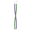
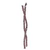
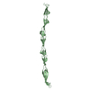
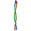
Journal: Proc Natl Acad Sci U S A / Year: 2006 Title: Crystal structures of human cardiac beta-myosin II S2-Delta provide insight into the functional role of the S2 subfragment. Authors: Wulf Blankenfeldt / Nicolas H Thomä / John S Wray / Mathias Gautel / Ilme Schlichting / Abstract: Myosin II is the major component of the muscle thick filament. It consists of two N-terminal S1 subfragments ("heads") connected to a long dimeric coiled-coil rod. The rod is in itself twofold ...Myosin II is the major component of the muscle thick filament. It consists of two N-terminal S1 subfragments ("heads") connected to a long dimeric coiled-coil rod. The rod is in itself twofold symmetric, but in the filament, the two heads point away from the filament surface and are therefore not equivalent. This breaking of symmetry requires the initial section of the rod, subfragment 2 (S2), to be relatively flexible. S2 is an important functional element, involved in various mechanisms by which the activity of smooth and striated muscle is regulated. We have determined crystal structures of the 126 N-terminal residues of S2 from human cardiac beta-myosin II (S2-Delta), of both WT and the disease-associated E924K mutant. S2-Delta is a straight parallel dimeric coiled coil, but the N terminus of one chain is disordered in WT-S2-Delta due to crystal contacts, indicative of unstable local structure. Bulky noncanonical side chains pack into a/d positions of S2-Delta's N terminus, leading to defined local asymmetry and axial stagger, which could induce nonequivalence of the S1 subfragments. Additionally, S2 possesses a conserved charge distribution with three prominent rings of negative potential within S2-Delta, the first of which may provide a binding interface for the "blocked head" of smooth muscle myosin in the OFF state. The observation that many disease-associated mutations affect the second negatively charged ring further suggests that charge interactions play an important role in regulation of cardiac muscle activity through myosin-binding protein C.
#1: Journal: J.Mol.Biol. / Year: 1999 Title: Mutations in Beta-Myosin S2 that Cause Familial Hypertrophic Cardiomyopathy (Fhc) Abolish the Interaction with the Regulatory Domain of Myosin-Binding Protein-C Authors: Gruen, M. / Gautel, M.
Resolution: 2.7→20 Å / Num. obs: 9756 / % possible obs: 96.7 % / Observed criterion σ(I): -3 / Redundancy: 5.7 % / Biso Wilson estimate: 53 Å2 / Rsym value: 0.104 / Net I/σ(I): 9.8
Reflection shell
Resolution: 2.7→2.8 Å / Redundancy: 3.4 % / Mean I/σ(I) obs: 3.1 / Rsym value: 0.326 / % possible all: 86.3
-
Processing
Software
Name
Version
Classification
XDS
datascaling
XDS
datareduction
SHELXD
phasing
SHARP
phasing
CNS
1
refinement
Refinement
Method to determine structure: MAD / Resolution: 2.7→20 Å / Isotropic thermal model: ISOTROPIC / Cross valid method: THROUGHOUT / Stereochemistry target values: Engh & Huber Details: Target sigma values for restrained B -factor refinement have been relaxed to 10 A*A to reflect the special shape of the molecule. Atoms that could not be located have been assigned a ...Details: Target sigma values for restrained B -factor refinement have been relaxed to 10 A*A to reflect the special shape of the molecule. Atoms that could not be located have been assigned a temperature factor of 500 A*A and occupancy value of 0 for better discernability.
Rfactor
Num. reflection
% reflection
Selection details
Rfree
0.283
496
5 %
RANDOM
Rwork
0.242
-
-
-
obs
0.242
9756
96.7 %
-
Displacement parameters
Biso mean: 77 Å2
Baniso -1
Baniso -2
Baniso -3
1-
-12.995 Å2
0 Å2
0 Å2
2-
-
-32.36 Å2
0 Å2
3-
-
-
45.354 Å2
Refinement step
Cycle: LAST / Resolution: 2.7→20 Å
Protein
Nucleic acid
Ligand
Solvent
Total
Num. atoms
1953
0
2
0
1955
Refine LS restraints
Refine-ID
Type
Dev ideal
X-RAY DIFFRACTION
c_bond_d
0.009
X-RAY DIFFRACTION
c_angle_deg
1.18
LS refinement shell
Resolution: 2.7→2.87 Å
Rfactor
Num. reflection
% reflection
Rfree
0.393
37
-
Rwork
0.353
-
-
obs
-
-
86.3 %
+
About Yorodumi
-
News
-
Feb 9, 2022. New format data for meta-information of EMDB entries
New format data for meta-information of EMDB entries
Version 3 of the EMDB header file is now the official format.
The previous official version 1.9 will be removed from the archive.
In the structure databanks used in Yorodumi, some data are registered as the other names, "COVID-19 virus" and "2019-nCoV". Here are the details of the virus and the list of structure data.
Jan 31, 2019. EMDB accession codes are about to change! (news from PDBe EMDB page)
EMDB accession codes are about to change! (news from PDBe EMDB page)
The allocation of 4 digits for EMDB accession codes will soon come to an end. Whilst these codes will remain in use, new EMDB accession codes will include an additional digit and will expand incrementally as the available range of codes is exhausted. The current 4-digit format prefixed with “EMD-” (i.e. EMD-XXXX) will advance to a 5-digit format (i.e. EMD-XXXXX), and so on. It is currently estimated that the 4-digit codes will be depleted around Spring 2019, at which point the 5-digit format will come into force.
The EM Navigator/Yorodumi systems omit the EMD- prefix.
Related info.:Q: What is EMD? / ID/Accession-code notation in Yorodumi/EM Navigator
Yorodumi is a browser for structure data from EMDB, PDB, SASBDB, etc.
This page is also the successor to EM Navigator detail page, and also detail information page/front-end page for Omokage search.
The word "yorodu" (or yorozu) is an old Japanese word meaning "ten thousand". "mi" (miru) is to see.
Related info.:EMDB / PDB / SASBDB / Comparison of 3 databanks / Yorodumi Search / Aug 31, 2016. New EM Navigator & Yorodumi / Yorodumi Papers / Jmol/JSmol / Function and homology information / Changes in new EM Navigator and Yorodumi
 Movie
Movie Controller


 Open data
 Basic information
 Components
 Keywords
 Function and homology information
 Homo sapiens (human)
 X-RAY DIFFRACTION /
  Authors
 Citation
 Structure visualization
 Downloads & links
 Other downloads
 PDBj
PDBj


 Assembly


 Mass: 200.590 Da / Num. of mol.: 2 / Source method: obtained synthetically / Formula: Hg
 Sample preparation
 / Beamline: ID29 / Wavelength: 1.0088, 0.82655
 Processing