[English] 日本語
Yorodumi
- PDB-4wve: Crystal structure of the Staphylococcus aureus SasG G52-E2-G53 module -

+
Open data


ID or keywords:

Loading...

-
Basic information

Entry
Database: PDB / ID: 4wve
TitleCrystal structure of the Staphylococcus aureus SasG G52-E2-G53 module
ComponentsSurface protein G
KeywordsSTRUCTURAL PROTEIN / single-layer beta sheet / biofilm formation / surface
Function / homology
Function and homology information


single-species submerged biofilm formation / extracellular region
Similarity search - Function
Resuscitation-promoting factor rpfb fold - #30 / Resuscitation-promoting factor rpfb fold / E domain / E domain / Bacterial lectin / G5 domain / G5 domain / G5 domain profile. / G5 / YSIRK type signal peptide ...Resuscitation-promoting factor rpfb fold - #30 / Resuscitation-promoting factor rpfb fold / E domain / E domain / Bacterial lectin / G5 domain / G5 domain / G5 domain profile. / G5 / YSIRK type signal peptide / YSIRK Gram-positive signal peptide / LPXTG cell wall anchor motif / Gram-positive cocci surface proteins LPxTG motif profile. / LPXTG cell wall anchor domain / Single Sheet / Mainly Beta
Similarity search - Domain/homology
Biological speciesStaphylococcus aureus (bacteria)
MethodX-RAY DIFFRACTION / SYNCHROTRON / MOLECULAR REPLACEMENT / molecular replacement / Resolution: 1.6 Å
AuthorsWhelan, F. / Potts, J.R.
Funding support United Kingdom, 1items
OrganizationGrant numberCountry
Biotechnology and Biological Sciences Research CouncilBB/J005029/1 United Kingdom
Citation
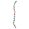
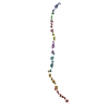
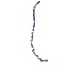
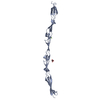
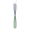
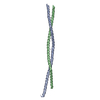
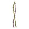
Journal: Nat Commun / Year: 2015
Title: Cooperative folding of intrinsically disordered domains drives assembly of a strong elongated protein.
Authors: Gruszka, D.T. / Whelan, F. / Farrance, O.E. / Fung, H.K. / Paci, E. / Jeffries, C.M. / Svergun, D.I. / Baldock, C. / Baumann, C.G. / Brockwell, D.J. / Potts, J.R. / Clarke, J.
History
DepositionNov 5, 2014Deposition site: RCSB / Processing site: PDBE
Revision 1.0Jun 3, 2015Provider: repository / Type: Initial release
Revision 1.1Jun 10, 2015Group: Database references
Revision 1.2Jun 17, 2015Group: Database references
Revision 2.0Aug 30, 2017Group: Atomic model / Author supporting evidence / Derived calculations
Category: atom_site / pdbx_audit_support / struct_site_gen
Item: _atom_site.B_iso_or_equiv / _atom_site.Cartn_x ..._atom_site.B_iso_or_equiv / _atom_site.Cartn_x / _atom_site.Cartn_y / _atom_site.Cartn_z / _atom_site.occupancy / _pdbx_audit_support.funding_organization / _struct_site_gen.auth_seq_id
Revision 2.1Jan 10, 2024Group: Data collection / Database references / Refinement description
Category: chem_comp_atom / chem_comp_bond ...chem_comp_atom / chem_comp_bond / database_2 / pdbx_initial_refinement_model / struct_ncs_dom_lim
Item: _database_2.pdbx_DOI / _database_2.pdbx_database_accession ..._database_2.pdbx_DOI / _database_2.pdbx_database_accession / _struct_ncs_dom_lim.beg_auth_comp_id / _struct_ncs_dom_lim.beg_label_asym_id / _struct_ncs_dom_lim.beg_label_comp_id / _struct_ncs_dom_lim.beg_label_seq_id / _struct_ncs_dom_lim.end_auth_comp_id / _struct_ncs_dom_lim.end_label_asym_id / _struct_ncs_dom_lim.end_label_comp_id / _struct_ncs_dom_lim.end_label_seq_id

-
Structure visualization

Structure viewerMolecule:
MolmilJmol/JSmol

Downloads & links

-
Assembly

Deposited unit
A: Surface protein G
B: Surface protein G
hetero molecules


Theoretical massNumber of molelcules
Total (without water)47,4193
Polymers47,3832
Non-polymers351
Water4,630257
1
A: Surface protein G
hetero molecules


Theoretical massNumber of molelcules
Total (without water)23,7272
Polymers23,6921
Non-polymers351
Water181
TypeNameSymmetry operationNumber
identity operation1_555x,y,z1
2
B: Surface protein G


Theoretical massNumber of molelcules
Total (without water)23,6921
Polymers23,6921
Non-polymers00
Water181
TypeNameSymmetry operationNumber
identity operation1_555x,y,z1
Unit cell
Length a, b, c (Å)39.861, 45.628, 60.359
Angle α, β, γ (deg.)84.760, 83.220, 78.600
Int Tables number1
Space group name H-MP1
Noncrystallographic symmetry (NCS)NCS domain:
IDEns-IDDetails
11A
21B

NCS domain segments:

Component-ID: 0 / Ens-ID: 1 / Beg auth comp-ID: GLY / Beg label comp-ID: GLY / End auth comp-ID: GLU / End label comp-ID: GLU / Refine code: 0 / Auth seq-ID: 552 - 756 / Label seq-ID: 10 - 214

Dom-IDAuth asym-IDLabel asym-ID
1AA
2BB

-
Components

#1: Protein Surface protein G /


Mass: 23691.529 Da / Num. of mol.: 2 / Fragment: UNP residues 545-757
Source method: isolated from a genetically manipulated source
Source: (gene. exp.) Staphylococcus aureus (bacteria) / Strain: NCTC 8325 / Gene: sasG, SAOUHSC_02798 / Plasmid: pSKB2 / Production host: Escherichia coli (E. coli) / Strain (production host): BL21(DE3) / References: UniProt: Q2G2B2
#2: Chemical ChemComp-CL / CHLORIDE ION / Chloride


Mass: 35.453 Da / Num. of mol.: 1 / Source method: obtained synthetically / Formula: Cl
#3: Water ChemComp-HOH / water / Water


Mass: 18.015 Da / Num. of mol.: 257 / Source method: isolated from a natural source / Formula: H2O

-
Experimental details

-
Experiment

ExperimentMethod: X-RAY DIFFRACTION / Number of used crystals: 1

-
Sample preparation

CrystalDensity Matthews: 2.25 Å3/Da / Density % sol: 45.33 %
Crystal growTemperature: 293 K / Method: vapor diffusion, sitting drop / pH: 7.5
Details: 0.2 M magnesium chloride, 0.1 M HEPES, 30% PEG400, pH 7.5

-
Data collection

DiffractionMean temperature: 100 K
Diffraction sourceSource: SYNCHROTRON / Site: Diamond / Beamline: I04 / Wavelength: 0.9795 Å
DetectorType: DECTRIS PILATUS 6M-F / Detector: PIXEL / Date: Feb 23, 2014
RadiationProtocol: SINGLE WAVELENGTH / Monochromatic (M) / Laue (L): M / Scattering type: x-ray
Radiation wavelengthWavelength: 0.9795 Å / Relative weight: 1
ReflectionResolution: 1.6→59.79 Å / Num. obs: 53868 / % possible obs: 98.9 % / Redundancy: 4.3 % / Biso Wilson estimate: 14.4 Å2 / CC1/2: 0.992 / Rmerge(I) obs: 0.089 / Rpim(I) all: 0.05 / Net I/σ(I): 15.8 / Num. measured all: 225293 / Scaling rejects: 717
Reflection shell

Diffraction-ID: 1 / Redundancy: 4.3 % / Rejects: 0

Resolution (Å)Rmerge(I) obsMean I/σ(I) obsNum. measured allNum. unique allCC1/2Rpim(I) all% possible all
1.6-1.630.5114.61126026310.9290.27897.9
8.76-59.790.0783914173280.990.04198.8

-
Phasing

PhasingMethod: molecular replacement

-
Processing

Software
NameVersionClassification
XDS0.2.17data reduction
Aimless0.2.17data scaling
REFMAC5.8.0073refinement
PDB_EXTRACT3.15data extraction
PHASERphasing
RefinementMethod to determine structure: MOLECULAR REPLACEMENT
Starting model: 3TIP
Resolution: 1.6→59.79 Å / Cor.coef. Fo:Fc: 0.957 / Cor.coef. Fo:Fc free: 0.934 / WRfactor Rfree: 0.2664 / WRfactor Rwork: 0.2244 / FOM work R set: 0.8058 / SU B: 4.482 / SU ML: 0.081 / SU R Cruickshank DPI: 0.1043 / SU Rfree: 0.1068 / Cross valid method: FREE R-VALUE / σ(F): 0 / ESU R: 0.104 / ESU R Free: 0.107 / Stereochemistry target values: MAXIMUM LIKELIHOOD
Details: HYDROGENS HAVE BEEN ADDED IN THE RIDING POSITIONS U VALUES : WITH TLS ADDED
RfactorNum. reflection% reflectionSelection details
Rfree0.249 2694 5 %RANDOM
Rwork0.2071 50959 --
obs0.2093 50959 98.47 %-
Solvent computationIon probe radii: 0.8 Å / Shrinkage radii: 0.8 Å / VDW probe radii: 1.2 Å / Solvent model: MASK
Displacement parametersBiso max: 56.9 Å2 / Biso mean: 25.569 Å2 / Biso min: 11.53 Å2
Baniso -1Baniso -2Baniso -3
1-1.79 Å20.42 Å20.05 Å2
2---0.28 Å21.2 Å2
3----1.14 Å2
Refinement stepCycle: final / Resolution: 1.6→59.79 Å
ProteinNucleic acidLigandSolventTotal
Num. atoms3170 0 1 257 3428
Biso mean--16.5 31.49 -
Num. residues----420
Refine LS restraints
Refine-IDTypeDev idealDev ideal targetNumber
X-RAY DIFFRACTIONr_bond_refined_d0.0140.023321
X-RAY DIFFRACTIONr_bond_other_d0.0050.023155
X-RAY DIFFRACTIONr_angle_refined_deg1.7252.0184531
X-RAY DIFFRACTIONr_angle_other_deg1.11537426
X-RAY DIFFRACTIONr_dihedral_angle_1_deg6.0535440
X-RAY DIFFRACTIONr_dihedral_angle_2_deg37.10226.803122
X-RAY DIFFRACTIONr_dihedral_angle_3_deg12.53915596
X-RAY DIFFRACTIONr_dihedral_angle_4_deg18.9021510
X-RAY DIFFRACTIONr_chiral_restr0.1010.2518
X-RAY DIFFRACTIONr_gen_planes_refined0.0080.0223692
X-RAY DIFFRACTIONr_gen_planes_other0.0040.02546
X-RAY DIFFRACTIONr_mcbond_it1.3461.461718
X-RAY DIFFRACTIONr_mcbond_other1.3471.461717
X-RAY DIFFRACTIONr_mcangle_it2.0072.1892157
Refine LS restraints NCS

Ens-ID: 1 / Number: 10074 / Refine-ID: X-RAY DIFFRACTION / Type: interatomic distance / Rms dev position: 0.14 Å / Weight position: 0.05

Dom-IDAuth asym-ID
1A
2B
LS refinement shell
Resolution (Å)Rfactor RfreeNum. reflection RfreeRfactor RworkNum. reflection RworkRefine-ID% reflection obs (%)
1.6-1.6420.2921850.2673686X-RAY DIFFRACTION96.27
1.642-1.6870.282250.2473575X-RAY DIFFRACTION96.643
1.687-1.7350.2861910.2343541X-RAY DIFFRACTION97.441
1.735-1.7890.291490.2263432X-RAY DIFFRACTION97.735
1.789-1.8470.2661640.2193372X-RAY DIFFRACTION97.68
1.847-1.9120.2411700.2163257X-RAY DIFFRACTION98.449
1.912-1.9840.2091580.2043158X-RAY DIFFRACTION98.31
1.984-2.0650.2691640.2182994X-RAY DIFFRACTION98.227
2.065-2.1570.2451610.2122915X-RAY DIFFRACTION98.97
2.157-2.2620.2541420.2092787X-RAY DIFFRACTION99.087
2.262-2.3840.2731410.2072664X-RAY DIFFRACTION99.398
2.384-2.5290.2361360.2082527X-RAY DIFFRACTION99.514
2.529-2.7030.2371180.2192356X-RAY DIFFRACTION99.678
2.703-2.9190.2741100.2282197X-RAY DIFFRACTION99.697
2.919-3.1970.2061120.2022033X-RAY DIFFRACTION99.721
3.197-3.5730.288990.2071842X-RAY DIFFRACTION99.949
3.573-4.1230.2631000.1871610X-RAY DIFFRACTION99.942
4.123-5.0440.195800.1651374X-RAY DIFFRACTION100
5.044-7.1070.243540.2061051X-RAY DIFFRACTION99.91
7.107-59.790.278350.21587X-RAY DIFFRACTION99.52
Refinement TLS params.

Method: refined / Refine-ID: X-RAY DIFFRACTION

IDL112)L122)L132)L222)L232)L332)S11 (Å °)S12 (Å °)S13 (Å °)S21 (Å °)S22 (Å °)S23 (Å °)S31 (Å °)S32 (Å °)S33 (Å °)T112)T122)T132)T222)T232)T332)Origin x (Å)Origin y (Å)Origin z (Å)
10.1744-0.52710.90331.9459-3.37155.8772-0.0012-0.0205-0.00640.10270.0262-0.0076-0.1784-0.0184-0.0250.1067-0.0057-0.02970.0493-0.01330.0607-4.2119-31.446148.512
20.0348-0.12150.27480.6245-1.42343.43460.049-0.0061-0.0138-0.07370.04820.04330.15490.0148-0.09720.113-0.0013-0.01590.0849-0.00120.038-6.5262-24.239824.9883
30.1340.2826-0.40981.5751-2.55764.19250.04850.00380.02850.106-0.05680.0176-0.13870.10910.00830.11710.03840.02510.05580.00680.0567-1.45227.3762-50.828
40.0548-0.32370.31352.0142-1.95911.9086-0.0037-0.0099-0.0107-0.076-0.00370.0070.07130.00910.00740.140.05960.01370.03990.03730.0705-27.5454-4.7672-2.3397
50.22310.4654-0.41376.3925-6.61746.888-0.0193-0.0327-0.028-0.2789-0.0634-0.05860.2820.05720.08260.07090.03690.02540.07260.01210.1049-24.1865-45.614932.4773
60.27250.6627-0.74812.8057-3.3714.39320.17160.03230.07710.00390.0340.30580.09320.2094-0.20570.21790.05970.02180.16120.1050.1003-27.6122-73.830662.7542
Refinement TLS group
IDRefine-IDRefine TLS-IDAuth asym-IDAuth seq-ID
1X-RAY DIFFRACTION1A543 - 588
2X-RAY DIFFRACTION2A589 - 677
3X-RAY DIFFRACTION3A678 - 757
4X-RAY DIFFRACTION4B551 - 651
5X-RAY DIFFRACTION5B652 - 694
6X-RAY DIFFRACTION6B695 - 757

+
About Yorodumi

-
News

-
Feb 9, 2022. New format data for meta-information of EMDB entries

New format data for meta-information of EMDB entries

  • Version 3 of the EMDB header file is now the official format.
  • The previous official version 1.9 will be removed from the archive.

Related info.:EMDB header

External links:wwPDB to switch to version 3 of the EMDB data model

-
Aug 12, 2020. Covid-19 info

Covid-19 info

URL: https://pdbj.org/emnavi/covid19.php

New page: Covid-19 featured information page in EM Navigator.

Related info.:Covid-19 info / Mar 5, 2020. Novel coronavirus structure data

+
Mar 5, 2020. Novel coronavirus structure data

Novel coronavirus structure data

Related info.:Yorodumi Speices / Aug 12, 2020. Covid-19 info

External links:COVID-19 featured content - PDBj / Molecule of the Month (242):Coronavirus Proteases

+
Jan 31, 2019. EMDB accession codes are about to change! (news from PDBe EMDB page)

EMDB accession codes are about to change! (news from PDBe EMDB page)

  • The allocation of 4 digits for EMDB accession codes will soon come to an end. Whilst these codes will remain in use, new EMDB accession codes will include an additional digit and will expand incrementally as the available range of codes is exhausted. The current 4-digit format prefixed with “EMD-” (i.e. EMD-XXXX) will advance to a 5-digit format (i.e. EMD-XXXXX), and so on. It is currently estimated that the 4-digit codes will be depleted around Spring 2019, at which point the 5-digit format will come into force.
  • The EM Navigator/Yorodumi systems omit the EMD- prefix.

Related info.:Q: What is EMD? / ID/Accession-code notation in Yorodumi/EM Navigator

External links:EMDB Accession Codes are Changing Soon! / Contact to PDBj

+
Jul 12, 2017. Major update of PDB

Major update of PDB

  • wwPDB released updated PDB data conforming to the new PDBx/mmCIF dictionary.
  • This is a major update changing the version number from 4 to 5, and with Remediation, in which all the entries are updated.
  • In this update, many items about electron microscopy experimental information are reorganized (e.g. em_software).
  • Now, EM Navigator and Yorodumi are based on the updated data.

External links:wwPDB Remediation / Enriched Model Files Conforming to OneDep Data Standards Now Available in the PDB FTP Archive

-
Yorodumi

Thousand views of thousand structures

  • Yorodumi is a browser for structure data from EMDB, PDB, SASBDB, etc.
  • This page is also the successor to EM Navigator detail page, and also detail information page/front-end page for Omokage search.
  • The word "yorodu" (or yorozu) is an old Japanese word meaning "ten thousand". "mi" (miru) is to see.

Related info.:EMDB / PDB / SASBDB / Comparison of 3 databanks / Yorodumi Search / Aug 31, 2016. New EM Navigator & Yorodumi / Yorodumi Papers / Jmol/JSmol / Function and homology information / Changes in new EM Navigator and Yorodumi

Read more