 Movie
Movie Controller

[English] 日本語
 Yorodumi
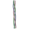
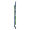
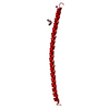
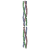
- PDB-6gaj: Crystal structure of the T1L reovirus sigma1 coiled coil tail (iodide) -
+ Open data

- Basic information
| Entry | Database: PDB / ID: 6gaj | ||||||
|---|---|---|---|---|---|---|---|
| Title | Crystal structure of the T1L reovirus sigma1 coiled coil tail (iodide) | ||||||
 Components Components | Outer capsid protein sigma-1 | ||||||
 Keywords Keywords | VIRAL PROTEIN / cell attachment protein / reovirus sigma1 / coiled coil | ||||||
| Function / homology |  Function and homology information Function and homology informationviral outer capsid / viral capsid / cell adhesion / symbiont entry into host cell / virion attachment to host cell Similarity search - Function | ||||||
| Biological species |  Mammalian orthoreovirus 1 Lang Mammalian orthoreovirus 1 Lang | ||||||
| Method |  X-RAY DIFFRACTION / SYNCHROTRON / SAD / Resolution: 1.35 Å X-RAY DIFFRACTION / SYNCHROTRON / SAD / Resolution: 1.35 Å | ||||||
 Authors Authors | Dietrich, M.H. / Stehle, T. | ||||||
| Funding support |  United States, 1items United States, 1items
| ||||||
 Citation Citation | Journal: J. Virol. / Year: 2018 Title: Structural and Functional Features of the Reovirus sigma 1 Tail. Authors: Dietrich, M.H. / Ogden, K.M. / Long, J.M. / Ebenhoch, R. / Thor, A. / Dermody, T.S. / Stehle, T. | ||||||
| History |
|
- Structure visualization
| Structure viewer | Molecule: MolmilJmol/JSmol |
|---|
- Downloads & links
-Download
| PDBx/mmCIF format | 6gaj.cif.gz | 107.8 KB | Display | PDBx/mmCIF format |
|---|---|---|---|---|
| PDB format | pdb6gaj.ent.gz | 82.8 KB | Display | PDB format |
| PDBx/mmJSON format | 6gaj.json.gz | Tree view | PDBx/mmJSON format | |
| Others |  Other downloads Other downloads |
-Validation report
| Arichive directory | https://data.pdbj.org/pub/pdb/validation_reports/ga/6gajftp://data.pdbj.org/pub/pdb/validation_reports/ga/6gaj | HTTPS FTP |
|---|
-Related structure data


-Links
 PDBj
PDBj
- Assembly
| Deposited unit | 
| ||||||||
|---|---|---|---|---|---|---|---|---|---|
| 1 |
| ||||||||
| Unit cell |
|
-Components
| #1: Protein | Mass: 14296.860 Da / Num. of mol.: 3 Source method: isolated from a genetically manipulated source Source: (gene. exp.) Mammalian orthoreovirus 1 Lang / Gene: S1 / Production host:  #2: Chemical |   Mass: 126.904 Da / Num. of mol.: 3 / Source method: obtained synthetically / Formula: I Mass: 126.904 Da / Num. of mol.: 3 / Source method: obtained synthetically / Formula: I#3: Chemical | ChemComp-CL /   Mass: 35.453 Da / Num. of mol.: 4 / Source method: obtained synthetically / Formula: Cl Mass: 35.453 Da / Num. of mol.: 4 / Source method: obtained synthetically / Formula: Cl#4: Water | ChemComp-HOH / |  Mass: 18.015 Da / Num. of mol.: 817 / Source method: isolated from a natural source / Formula: H2O Mass: 18.015 Da / Num. of mol.: 817 / Source method: isolated from a natural source / Formula: H2O |
|---|
-Experimental details
-Experiment
| Experiment | Method: X-RAY DIFFRACTION / Number of used crystals: 1 |
|---|
- Sample preparation
| Crystal | Density Matthews: 2.04 Å3/Da / Density % sol: 40.59 % |
|---|---|
| Crystal grow | Temperature: 277 K / Method: vapor diffusion, sitting drop / pH: 9.5 / Details: 18% (v/v) PEG8000, 0.1M CAPS pH 9.5, 0.2M NaI |
-Data collection
| Diffraction | Mean temperature: 100 K |
|---|---|
| Diffraction source | Source: SYNCHROTRON / Site: SLS  / Beamline: X06DA / Wavelength: 1 Å / Beamline: X06DA / Wavelength: 1 Å |
| Detector | Type: DECTRIS PILATUS 2M-F / Detector: PIXEL / Date: May 24, 2014 |
| Radiation | Protocol: SINGLE WAVELENGTH / Monochromatic (M) / Laue (L): M / Scattering type: x-ray |
| Radiation wavelength | Wavelength: 1 Å / Relative weight: 1 |
| Reflection | Resolution: 1.35→48.73 Å / Num. obs: 76273 / % possible obs: 99.9 % / Redundancy: 6.6 % / Biso Wilson estimate: 12.04 Å2 / CC1/2: 0.999 / Rrim(I) all: 0.109 / Net I/σ(I): 12 |
| Reflection shell | Resolution: 1.35→1.39 Å / Redundancy: 6 % / Mean I/σ(I) obs: 1.9 / Num. unique obs: 5563 / CC1/2: 0.723 / Rrim(I) all: 0.876 / % possible all: 99.9 |
- Processing
| Software |
| ||||||||||||||||||||||||||||||||||||||||||||||||||||||||||||||||||||||||||||||||||||||||||||||||||||||||||||||||||
|---|---|---|---|---|---|---|---|---|---|---|---|---|---|---|---|---|---|---|---|---|---|---|---|---|---|---|---|---|---|---|---|---|---|---|---|---|---|---|---|---|---|---|---|---|---|---|---|---|---|---|---|---|---|---|---|---|---|---|---|---|---|---|---|---|---|---|---|---|---|---|---|---|---|---|---|---|---|---|---|---|---|---|---|---|---|---|---|---|---|---|---|---|---|---|---|---|---|---|---|---|---|---|---|---|---|---|---|---|---|---|---|---|---|---|---|
| Refinement | Method to determine structure: SAD / Resolution: 1.35→48.73 Å / Cor.coef. Fo:Fc: 0.9488 / Cor.coef. Fo:Fc free: 0.9443 / SU R Cruickshank DPI: 0.06 / Cross valid method: THROUGHOUT / σ(F): 0 / SU R Blow DPI: 0.067 / SU Rfree Blow DPI: 0.067 / SU Rfree Cruickshank DPI: 0.062
| ||||||||||||||||||||||||||||||||||||||||||||||||||||||||||||||||||||||||||||||||||||||||||||||||||||||||||||||||||
| Displacement parameters | Biso mean: 15.97 Å2
| ||||||||||||||||||||||||||||||||||||||||||||||||||||||||||||||||||||||||||||||||||||||||||||||||||||||||||||||||||
| Refine analyze | Luzzati coordinate error obs: 0.148 Å | ||||||||||||||||||||||||||||||||||||||||||||||||||||||||||||||||||||||||||||||||||||||||||||||||||||||||||||||||||
| Refinement step | Cycle: 1 / Resolution: 1.35→48.73 Å
| ||||||||||||||||||||||||||||||||||||||||||||||||||||||||||||||||||||||||||||||||||||||||||||||||||||||||||||||||||
| Refine LS restraints |
| ||||||||||||||||||||||||||||||||||||||||||||||||||||||||||||||||||||||||||||||||||||||||||||||||||||||||||||||||||
| LS refinement shell | Resolution: 1.35→1.39 Å / Total num. of bins used: 20
|
