ムービー
ムービー コントローラー


+ データを開く

- 基本情報
基本情報
| 登録情報 | データベース: EMDB / ID: EMD-5779 | |||||||||
|---|---|---|---|---|---|---|---|---|---|---|
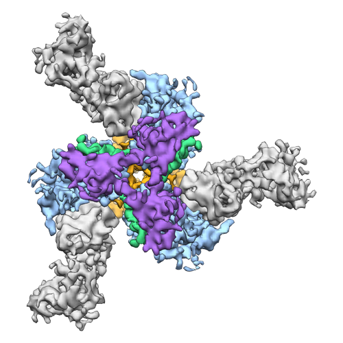
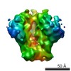
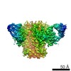
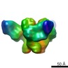
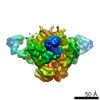
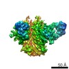
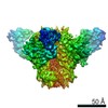
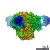
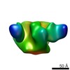
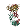
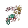
| タイトル | Cryo-EM structure of the BG505 SOSIP.664 HIV-1 Env trimer with 3 PGV04 Fabs | |||||||||
 マップデータ マップデータ | Cryo-EM structure of a fully glycosylated SOSIP.664 Env trimer with 3 broadly neutralizing PGV04 Fabs | |||||||||
 試料 試料 |
| |||||||||
 キーワード キーワード | HIV-1 trimeric spike / gp140 / SOSIP /  broadly neutralizing antibody (中和抗体) / PGV04 / Env / Envelope glycoprotein broadly neutralizing antibody (中和抗体) / PGV04 / Env / Envelope glycoprotein | |||||||||
| 機能・相同性 |  機能・相同性情報 機能・相同性情報positive regulation of plasma membrane raft polarization / positive regulation of receptor clustering / positive regulation of establishment of T cell polarity / virus-mediated perturbation of host defense response / host cell endosome membrane / clathrin-dependent endocytosis of virus by host cell / viral protein processing / fusion of virus membrane with host plasma membrane / fusion of virus membrane with host endosome membrane / エンベロープ (ウイルス) ...positive regulation of plasma membrane raft polarization / positive regulation of receptor clustering / positive regulation of establishment of T cell polarity / virus-mediated perturbation of host defense response / host cell endosome membrane / clathrin-dependent endocytosis of virus by host cell / viral protein processing / fusion of virus membrane with host plasma membrane / fusion of virus membrane with host endosome membrane / エンベロープ (ウイルス) / virion attachment to host cell / host cell plasma membrane / virion membrane / structural molecule activity / identical protein binding / 細胞膜類似検索 - 分子機能 | |||||||||
| 生物種 |   Human immunodeficiency virus 1 (ヒト免疫不全ウイルス) / Human immunodeficiency virus 1 (ヒト免疫不全ウイルス) /  Homo sapiens (ヒト) Homo sapiens (ヒト) | |||||||||
| 手法 | 単粒子再構成法 / クライオ電子顕微鏡法 / 解像度: 5.8 Å | |||||||||
 データ登録者 データ登録者 | Lyumkis D / Julien JP / de Val N / Cupo A / Potter CS / Klasse PJ / Burton DR / Sanders RW / Moore JP / Carragher B ...Lyumkis D / Julien JP / de Val N / Cupo A / Potter CS / Klasse PJ / Burton DR / Sanders RW / Moore JP / Carragher B / Wilson IA / Ward AB | |||||||||
 引用 引用 | ジャーナル: Science / 年: 2013 タイトル: Cryo-EM structure of a fully glycosylated soluble cleaved HIV-1 envelope trimer. 著者: Dmitry Lyumkis / Jean-Philippe Julien / Natalia de Val / Albert Cupo / Clinton S Potter / Per-Johan Klasse / Dennis R Burton / Rogier W Sanders / John P Moore / Bridget Carragher / Ian A Wilson / Andrew B Ward /  要旨: The HIV-1 envelope glycoprotein (Env) trimer contains the receptor binding sites and membrane fusion machinery that introduce the viral genome into the host cell. As the only target for broadly ...The HIV-1 envelope glycoprotein (Env) trimer contains the receptor binding sites and membrane fusion machinery that introduce the viral genome into the host cell. As the only target for broadly neutralizing antibodies (bnAbs), Env is a focus for rational vaccine design. We present a cryo-electron microscopy reconstruction and structural model of a cleaved, soluble Env trimer (termed BG505 SOSIP.664 gp140) in complex with a CD4 binding site (CD4bs) bnAb, PGV04, at 5.8 angstrom resolution. The structure reveals the spatial arrangement of Env components, including the V1/V2, V3, HR1, and HR2 domains, as well as shielding glycans. The structure also provides insights into trimer assembly, gp120-gp41 interactions, and the CD4bs epitope cluster for bnAbs, which covers a more extensive area and defines a more complex site of vulnerability than previously described. | |||||||||
| 履歴 |
|
- 構造の表示
構造の表示
| ムービー |
ムービービューア |
|---|---|
| 構造ビューア | EMマップ: SurfViewMolmilJmol/JSmol |
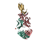
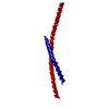
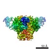
| 添付画像 |
- ダウンロードとリンク
-EMDBアーカイブ
| マップデータ | emd_5779.map.gz | 56.7 MB | EMDBマップデータ形式 | |
|---|---|---|---|---|
| ヘッダ (付随情報) | emd-5779-v30.xmlemd-5779.xml | 13.7 KB 13.7 KB | 表示 表示 | EMDBヘッダ |
| 画像 |  emd_5779_1.png emd_5779_1.png | 175.8 KB | ||
| アーカイブディレクトリ |  http://ftp.pdbj.org/pub/emdb/structures/EMD-5779ftp://ftp.pdbj.org/pub/emdb/structures/EMD-5779 http://ftp.pdbj.org/pub/emdb/structures/EMD-5779ftp://ftp.pdbj.org/pub/emdb/structures/EMD-5779 | HTTPS FTP |
-関連構造データ

-リンク
| EMDBのページ | EMDB (EBI/PDBe) / EMDataResource |
|---|---|
| 「今月の分子」の関連する項目 |


-マップ
| ファイル | ダウンロード / ファイル: emd_5779.map.gz / 形式: CCP4 / 大きさ: 62.5 MB / タイプ: IMAGE STORED AS FLOATING POINT NUMBER (4 BYTES) | ||||||||||||||||||||||||||||||||||||||||||||||||||||||||||||||||||||
|---|---|---|---|---|---|---|---|---|---|---|---|---|---|---|---|---|---|---|---|---|---|---|---|---|---|---|---|---|---|---|---|---|---|---|---|---|---|---|---|---|---|---|---|---|---|---|---|---|---|---|---|---|---|---|---|---|---|---|---|---|---|---|---|---|---|---|---|---|---|
| 注釈 | Cryo-EM structure of a fully glycosylated SOSIP.664 Env trimer with 3 broadly neutralizing PGV04 Fabs | ||||||||||||||||||||||||||||||||||||||||||||||||||||||||||||||||||||
| ボクセルのサイズ | X=Y=Z: 1.21 Å | ||||||||||||||||||||||||||||||||||||||||||||||||||||||||||||||||||||
| 密度 |
| ||||||||||||||||||||||||||||||||||||||||||||||||||||||||||||||||||||
| 対称性 | 空間群: 1 | ||||||||||||||||||||||||||||||||||||||||||||||||||||||||||||||||||||
| 詳細 | EMDB XML:
CCP4マップ ヘッダ情報:
| ||||||||||||||||||||||||||||||||||||||||||||||||||||||||||||||||||||
-添付データ
- 試料の構成要素
-全体 : Fully glycosylated BG505 SOSIP.664 Envelope trimer with 3 broadly...
| 全体 | 名称: Fully glycosylated BG505 SOSIP.664 Envelope trimer with 3 broadly neutralizing PGV04 Fabs |
|---|---|
| 要素 |
|
-超分子 #1000: Fully glycosylated BG505 SOSIP.664 Envelope trimer with 3 broadly...
| 超分子 | 名称: Fully glycosylated BG505 SOSIP.664 Envelope trimer with 3 broadly neutralizing PGV04 Fabs タイプ: sample / ID: 1000 集合状態: three SOSIP.664 gp140 subunits (trimeric HIV-1 spike) with 3 PGV04 Fabs Number unique components: 2 |
|---|---|
| 分子量 | 理論値: 600 KDa |
-分子 #1: BG505 SOSIP.664 HIV-1 Envelope glycoprotein gp140
| 分子 | 名称: BG505 SOSIP.664 HIV-1 Envelope glycoprotein gp140 / タイプ: protein_or_peptide / ID: 1 / Name.synonym: Env / コピー数: 3 / 集合状態: trimer / 組換発現: Yes |
|---|---|
| 由来(天然) | 生物種: Human immunodeficiency virus 1 (ヒト免疫不全ウイルス) 株: BG505.W6M.ENV.A5 / 別称: human immunodeficiency virus type I |
| 分子量 | 理論値: 600 KDa |
| 組換発現 | 生物種: Homo sapiens (ヒト) / 組換細胞: HEK 293T |
-分子 #2: Fragment antigen binding
| 分子 | 名称: Fragment antigen binding / タイプ: protein_or_peptide / ID: 2 / Name.synonym: Fab / コピー数: 3 / 集合状態: heterodimer / 組換発現: Yes |
|---|---|
| 由来(天然) | 生物種: Homo sapiens (ヒト) / 別称: Human |
| 分子量 | 実験値: 50 KDa / 理論値: 50 KDa |
| 組換発現 | 生物種: Homo sapiens (ヒト) / 組換細胞: HEK 293F |
-実験情報
-構造解析
| 手法 | クライオ電子顕微鏡法 |
|---|---|
 解析 解析 | 単粒子再構成法 |
| 試料の集合状態 | particle |
-試料調製
| 濃度 | 0.72 mg/mL |
|---|---|
| 緩衝液 | pH: 7.6 / 詳細: 20 mM Tris, 150 mM NaCl, 0.085 mM DDM |
| グリッド | 詳細: 400 mesh C-Flat CF-22-4C, plasma treated for 5 seconds |
| 凍結 | 凍結剤: ETHANE / 装置: HOMEMADE PLUNGER 手法: Specimen was prepared for cryo-EM by applying 3 microliters of sample to a freshly plasma cleaned holey carbon C-flat grid (Protochips, Inc.), allowing the sample to adsorb to the grid for 30 ...手法: Specimen was prepared for cryo-EM by applying 3 microliters of sample to a freshly plasma cleaned holey carbon C-flat grid (Protochips, Inc.), allowing the sample to adsorb to the grid for 30 seconds, followed by blotting with a small piece of filter paper and plunge-freezing into liquid ethane using a manual cryo-plunger in an ambient environment (4 degrees C). |
- 電子顕微鏡法
| 顕微鏡 | FEI TECNAI F20 |
|---|---|
| 電子線 | 加速電圧: 200 kV / 電子線源: FIELD EMISSION GUN |
| 電子光学系 | 倍率(補正後): 29000 / 照射モード: FLOOD BEAM / 撮影モード: BRIGHT FIELDBright-field microscopy / Cs: 2 mm / 最大 デフォーカス(公称値): 5.0 µm / 最小 デフォーカス(公称値): 1.0 µm / 倍率(公称値): 29000 |
| 試料ステージ | 試料ホルダーモデル: SIDE ENTRY, EUCENTRIC |
| アライメント法 | Legacy - 非点収差: objective lens astigmatism was corrected by observing Thon rings with the Leginon software |
| 詳細 | electron counting mode |
| 日付 | 2013年2月21日 |
| 撮影 | カテゴリ: CCD / フィルム・検出器のモデル: GATAN K2 (4k x 4k) / デジタル化 - サンプリング間隔: 5.0 µm / 実像数: 6355 / 平均電子線量: 32 e/Å2 詳細: The dose was fractionated over 20 raw frames collected over a 5-second exposure time (250 ms per frame) on the Gatan K2 Summit direct detection device, with each frame receiving a dose of ~9. ...詳細: The dose was fractionated over 20 raw frames collected over a 5-second exposure time (250 ms per frame) on the Gatan K2 Summit direct detection device, with each frame receiving a dose of ~9.4 e-/pixel/sec. The individual frames were aligned using a GPU-enabled frame-alignment program that was generously provided by Yifan Cheng and Xueming Li. This program was used to track the global shifts between individual frames. |
| Tilt angle min | 0 |
| 実験機器 |  モデル: Tecnai F20 / 画像提供: FEI Company |
-画像解析
| CTF補正 | 詳細: Frealign |
|---|---|
| 最終 再構成 | アルゴリズム: OTHER / 解像度のタイプ: BY AUTHOR / 解像度: 5.8 Å / 解像度の算出法: FSC 0.143 CUT-OFF / ソフトウェア - 名称: Xmipp, Frealign 詳細: Final maps were calculated after sorting for the presence of sub-stoichiometrically labeled trimers. 使用した粒子像数: 49572 |
| 詳細 | Reconstructed using resolution-limited refinement procedure implemented in Xmipp and Frealign. |
-原子モデル構築 1
| 初期モデル | PDB ID: |
|---|---|
| 精密化 | 空間: REAL |
| 得られたモデル |  PDB-3j5m: |
-原子モデル構築 2
| 初期モデル | PDB ID: |
|---|---|
| 精密化 | 空間: REAL |
| 得られたモデル | PDB-3j5m: |
-原子モデル構築 3
| 初期モデル | PDB ID: |
|---|---|
| 精密化 | 空間: REAL |
| 得られたモデル | PDB-3j5m: |