 Movie
Movie Controller

[English] 日本語
 Yorodumi
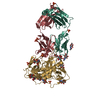
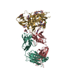
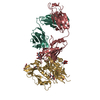
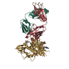
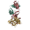
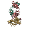
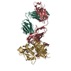
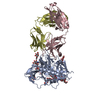
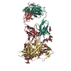
- PDB-3se9: Crystal structure of broadly and potently neutralizing antibody V... -
+ Open data

- Basic information
| Entry | Database: PDB / ID: 3se9 | ||||||
|---|---|---|---|---|---|---|---|
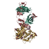
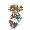
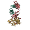
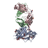
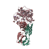
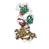
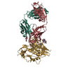
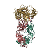
| Title | Crystal structure of broadly and potently neutralizing antibody VRC-PG04 in complex with HIV-1 gp120 | ||||||
 Components Components |
| ||||||
 Keywords Keywords | VIRAL PROTEIN/IMMUNE SYSTEM / HIV / GP120 / ANTIBODY / VRC-PG04 / NEUTRALIZATION / VACCINE / ENVELOPE GLYCOPROTEIN / VIRAL PROTEIN-IMMUNE SYSTEM COMPLEX | ||||||
| Function / homology |  Function and homology information Function and homology informationsymbiont-mediated perturbation of host defense response / positive regulation of plasma membrane raft polarization / positive regulation of receptor clustering / host cell endosome membrane / clathrin-dependent endocytosis of virus by host cell / viral protein processing / fusion of virus membrane with host plasma membrane / fusion of virus membrane with host endosome membrane / viral envelope / virion attachment to host cell ...symbiont-mediated perturbation of host defense response / positive regulation of plasma membrane raft polarization / positive regulation of receptor clustering / host cell endosome membrane / clathrin-dependent endocytosis of virus by host cell / viral protein processing / fusion of virus membrane with host plasma membrane / fusion of virus membrane with host endosome membrane / viral envelope / virion attachment to host cell / host cell plasma membrane / virion membrane / structural molecule activity / membrane Similarity search - Function | ||||||
| Biological species |   Human immunodeficiency virus 1 Human immunodeficiency virus 1 Homo sapiens (human) Homo sapiens (human) | ||||||
| Method |  X-RAY DIFFRACTION / SYNCHROTRON / MOLECULAR REPLACEMENT / Resolution: 2 Å X-RAY DIFFRACTION / SYNCHROTRON / MOLECULAR REPLACEMENT / Resolution: 2 Å | ||||||
 Authors Authors | Kwong, P.D. / Zhou, T. | ||||||
 Citation Citation | Journal: Science / Year: 2011 Title: Focused evolution of HIV-1 neutralizing antibodies revealed by structures and deep sequencing. Authors: Wu, X. / Zhou, T. / Zhu, J. / Zhang, B. / Georgiev, I. / Wang, C. / Chen, X. / Longo, N.S. / Louder, M. / McKee, K. / O'Dell, S. / Perfetto, S. / Schmidt, S.D. / Shi, W. / Wu, L. / Yang, Y. ...Authors: Wu, X. / Zhou, T. / Zhu, J. / Zhang, B. / Georgiev, I. / Wang, C. / Chen, X. / Longo, N.S. / Louder, M. / McKee, K. / O'Dell, S. / Perfetto, S. / Schmidt, S.D. / Shi, W. / Wu, L. / Yang, Y. / Yang, Z.Y. / Yang, Z. / Zhang, Z. / Bonsignori, M. / Crump, J.A. / Kapiga, S.H. / Sam, N.E. / Haynes, B.F. / Simek, M. / Burton, D.R. / Koff, W.C. / Doria-Rose, N.A. / Connors, M. / Mullikin, J.C. / Nabel, G.J. / Roederer, M. / Shapiro, L. / Kwong, P.D. / Mascola, J.R. | ||||||
| History |
|
- Structure visualization
| Structure viewer | Molecule: MolmilJmol/JSmol |
|---|
- Downloads & links
-Download
| PDBx/mmCIF format | 3se9.cif.gz | 464 KB | Display | PDBx/mmCIF format |
|---|---|---|---|---|
| PDB format | pdb3se9.ent.gz | 385.6 KB | Display | PDB format |
| PDBx/mmJSON format | 3se9.json.gz | Tree view | PDBx/mmJSON format | |
| Others |  Other downloads Other downloads |
-Validation report
| Arichive directory | https://data.pdbj.org/pub/pdb/validation_reports/se/3se9ftp://data.pdbj.org/pub/pdb/validation_reports/se/3se9 | HTTPS FTP |
|---|
-Related structure data


-Links
 PDBj
PDBj


- Assembly
| Deposited unit | 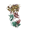
| ||||||||
|---|---|---|---|---|---|---|---|---|---|
| 1 |
| ||||||||
| Unit cell |
|
-Components
-Antibody , 2 types, 2 molecules HL
| #2: Antibody | Mass: 24644.771 Da / Num. of mol.: 1 / Fragment: Antigen binding fragement of heavy chain Source method: isolated from a genetically manipulated source Details: Co-transfection of both heavy and light chain plasmids Source: (gene. exp.) Homo sapiens (human) / Gene: antibody VRC-PG04 heavy chain / Plasmid: CMVR derived plasmids for IgG1 / Cell line (production host): HEK293F / Production host: Homo sapiens (human) |
|---|---|
| #3: Antibody | Mass: 23073.822 Da / Num. of mol.: 1 / Fragment: antibody light chain Source method: isolated from a genetically manipulated source Details: Co-transfection of both heavy and light chain plasmids Source: (gene. exp.) Homo sapiens (human) / Gene: antibody VRC-PG04 light chain / Plasmid: CMVR derived plasmids for IgG1 / Cell line (production host): HEK293F / Production host: Homo sapiens (human) |
-Protein / Sugars , 2 types, 11 molecules G
| #1: Protein | Mass: 39211.434 Da / Num. of mol.: 1 / Fragment: Core gp120 with trancations Source method: isolated from a genetically manipulated source Source: (gene. exp.) Human immunodeficiency virus 1 / Strain: 93TH057 / Gene: gp120 / Plasmid: pVRC8400 / Cell line (production host): HEK293F / Production host: Homo sapiens (human) / References: UniProt: Q0ED31*PLUS |
|---|---|
| #4: Sugar | ChemComp-NAG /  Type: D-saccharide, beta linking / Mass: 221.208 Da / Num. of mol.: 10 Type: D-saccharide, beta linking / Mass: 221.208 Da / Num. of mol.: 10Source method: isolated from a genetically manipulated source Formula: C8H15NO6 |
-Non-polymers , 6 types, 584 molecules 


| #5: Chemical | ChemComp-SO4 /  Mass: 96.063 Da / Num. of mol.: 5 / Source method: obtained synthetically / Formula: SO4 Mass: 96.063 Da / Num. of mol.: 5 / Source method: obtained synthetically / Formula: SO4#6: Chemical | ChemComp-GOL /  Mass: 92.094 Da / Num. of mol.: 4 / Source method: obtained synthetically / Formula: C3H8O3 Mass: 92.094 Da / Num. of mol.: 4 / Source method: obtained synthetically / Formula: C3H8O3#7: Chemical | ChemComp-CL /  Mass: 35.453 Da / Num. of mol.: 18 / Source method: obtained synthetically / Formula: Cl Mass: 35.453 Da / Num. of mol.: 18 / Source method: obtained synthetically / Formula: Cl#8: Chemical | ChemComp-BU3 / ( |  Mass: 90.121 Da / Num. of mol.: 1 / Source method: obtained synthetically / Formula: C4H10O2 Mass: 90.121 Da / Num. of mol.: 1 / Source method: obtained synthetically / Formula: C4H10O2#9: Chemical | ChemComp-TRS / |  Mass: 122.143 Da / Num. of mol.: 1 / Source method: obtained synthetically / Formula: C4H12NO3 / Comment: pH buffer*YM Mass: 122.143 Da / Num. of mol.: 1 / Source method: obtained synthetically / Formula: C4H12NO3 / Comment: pH buffer*YM#10: Water | ChemComp-HOH / | Mass: 18.015 Da / Num. of mol.: 555 / Source method: isolated from a natural source / Formula: H2O |
|---|
-Details
| Has protein modification | Y |
|---|
-Experimental details
-Experiment
| Experiment | Method: X-RAY DIFFRACTION / Number of used crystals: 1 |
|---|
- Sample preparation
| Crystal | Density Matthews: 2.8 Å3/Da / Density % sol: 56.13 % |
|---|---|
| Crystal grow | Temperature: 293 K / Method: vapor diffusion, hanging drop / pH: 7.5 Details: 9.9% PEG 4000, 9.0 % isopropanol, 100 mM Li2SO4, 100 mM HEPES, pH 7.5, VAPOR DIFFUSION, HANGING DROP, temperature 293K |
-Data collection
| Diffraction | Mean temperature: 100 K |
|---|---|
| Diffraction source | Source: SYNCHROTRON / Site: APS  / Beamline: 22-ID / Wavelength: 1 Å / Beamline: 22-ID / Wavelength: 1 Å |
| Detector | Type: MARMOSAIC 300 mm CCD / Detector: CCD / Date: Jun 18, 2010 |
| Radiation | Protocol: SINGLE WAVELENGTH / Monochromatic (M) / Laue (L): M / Scattering type: x-ray |
| Radiation wavelength | Wavelength: 1 Å / Relative weight: 1 |
| Reflection | Resolution: 2→50 Å / Num. all: 67230 / Num. obs: 59364 / % possible obs: 88.3 % |
- Processing
| Software |
| |||||||||||||||||||||||||||||||||||||||||||||||||||||||||||||||||||||||||||||||||||||||||||||||||||||||||||||||||||||||||||||||||||||||||||||||||||||||||||||||||||||||||||||||
|---|---|---|---|---|---|---|---|---|---|---|---|---|---|---|---|---|---|---|---|---|---|---|---|---|---|---|---|---|---|---|---|---|---|---|---|---|---|---|---|---|---|---|---|---|---|---|---|---|---|---|---|---|---|---|---|---|---|---|---|---|---|---|---|---|---|---|---|---|---|---|---|---|---|---|---|---|---|---|---|---|---|---|---|---|---|---|---|---|---|---|---|---|---|---|---|---|---|---|---|---|---|---|---|---|---|---|---|---|---|---|---|---|---|---|---|---|---|---|---|---|---|---|---|---|---|---|---|---|---|---|---|---|---|---|---|---|---|---|---|---|---|---|---|---|---|---|---|---|---|---|---|---|---|---|---|---|---|---|---|---|---|---|---|---|---|---|---|---|---|---|---|---|---|---|---|---|
| Refinement | Method to determine structure: MOLECULAR REPLACEMENT / Resolution: 2→23.734 Å / SU ML: 0.51 / σ(F): 1.93 / Phase error: 25.5 / Stereochemistry target values: ML
| |||||||||||||||||||||||||||||||||||||||||||||||||||||||||||||||||||||||||||||||||||||||||||||||||||||||||||||||||||||||||||||||||||||||||||||||||||||||||||||||||||||||||||||||
| Solvent computation | Shrinkage radii: 0.89 Å / VDW probe radii: 1 Å / Solvent model: FLAT BULK SOLVENT MODEL / Bsol: 30.269 Å2 / ksol: 0.349 e/Å3 | |||||||||||||||||||||||||||||||||||||||||||||||||||||||||||||||||||||||||||||||||||||||||||||||||||||||||||||||||||||||||||||||||||||||||||||||||||||||||||||||||||||||||||||||
| Displacement parameters |
| |||||||||||||||||||||||||||||||||||||||||||||||||||||||||||||||||||||||||||||||||||||||||||||||||||||||||||||||||||||||||||||||||||||||||||||||||||||||||||||||||||||||||||||||
| Refinement step | Cycle: LAST / Resolution: 2→23.734 Å
| |||||||||||||||||||||||||||||||||||||||||||||||||||||||||||||||||||||||||||||||||||||||||||||||||||||||||||||||||||||||||||||||||||||||||||||||||||||||||||||||||||||||||||||||
| Refine LS restraints |
| |||||||||||||||||||||||||||||||||||||||||||||||||||||||||||||||||||||||||||||||||||||||||||||||||||||||||||||||||||||||||||||||||||||||||||||||||||||||||||||||||||||||||||||||
| LS refinement shell |
| |||||||||||||||||||||||||||||||||||||||||||||||||||||||||||||||||||||||||||||||||||||||||||||||||||||||||||||||||||||||||||||||||||||||||||||||||||||||||||||||||||||||||||||||
| Refinement TLS params. | Method: refined / Refine-ID: X-RAY DIFFRACTION
| |||||||||||||||||||||||||||||||||||||||||||||||||||||||||||||||||||||||||||||||||||||||||||||||||||||||||||||||||||||||||||||||||||||||||||||||||||||||||||||||||||||||||||||||
| Refinement TLS group |
|
