 Movie
Movie Controller

[English] 日本語
 Yorodumi
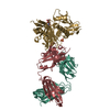
- PDB-6p8m: Crystal Structure of Antibody P-p3b3 A60C Heavy Chain in Complex ... -
+ Open data

- Basic information
| Entry | Database: PDB / ID: 6p8m | |||||||||
|---|---|---|---|---|---|---|---|---|---|---|
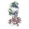
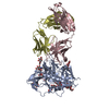
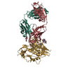
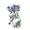
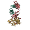
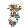
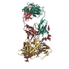
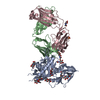
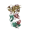
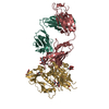
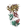
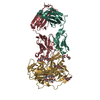
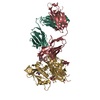
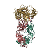
| Title | Crystal Structure of Antibody P-p3b3 A60C Heavy Chain in Complex with 426c HIV-1 gp120 core G459C | |||||||||
 Components Components |
| |||||||||
 Keywords Keywords | IMMUNE SYSTEM / antibody / 426c / immunization | |||||||||
| Function / homology |  Function and homology information Function and homology informationmembrane fusion involved in viral entry into host cell / host cell endosome membrane / viral envelope / symbiont entry into host cell / virion attachment to host cell / host cell plasma membrane / virion membrane / structural molecule activity Similarity search - Function | |||||||||
| Biological species |   Human immunodeficiency virus Human immunodeficiency virus | |||||||||
| Method |  X-RAY DIFFRACTION / SYNCHROTRON / MOLECULAR REPLACEMENT / Resolution: 3.594 Å X-RAY DIFFRACTION / SYNCHROTRON / MOLECULAR REPLACEMENT / Resolution: 3.594 Å | |||||||||
 Authors Authors | Weidle, C. / Pancera, M. | |||||||||
| Funding support |  United States, 1items United States, 1items
| |||||||||
 Citation Citation | Journal: Cell Rep / Year: 2019 Title: Overcoming Steric Restrictions of VRC01 HIV-1 Neutralizing Antibodies through Immunization. Authors: Parks, K.R. / MacCamy, A.J. / Trichka, J. / Gray, M. / Weidle, C. / Borst, A.J. / Khechaduri, A. / Takushi, B. / Agrawal, P. / Guenaga, J. / Wyatt, R.T. / Coler, R. / Seaman, M. / LaBranche, ...Authors: Parks, K.R. / MacCamy, A.J. / Trichka, J. / Gray, M. / Weidle, C. / Borst, A.J. / Khechaduri, A. / Takushi, B. / Agrawal, P. / Guenaga, J. / Wyatt, R.T. / Coler, R. / Seaman, M. / LaBranche, C. / Montefiori, D.C. / Veesler, D. / Pancera, M. / McGuire, A. / Stamatatos, L. | |||||||||
| History |
|
- Structure visualization
| Structure viewer | Molecule: MolmilJmol/JSmol |
|---|
- Downloads & links
-Download
| PDBx/mmCIF format | 6p8m.cif.gz | 289 KB | Display | PDBx/mmCIF format |
|---|---|---|---|---|
| PDB format | pdb6p8m.ent.gz | 232 KB | Display | PDB format |
| PDBx/mmJSON format | 6p8m.json.gz | Tree view | PDBx/mmJSON format | |
| Others |  Other downloads Other downloads |
-Validation report
| Arichive directory | https://data.pdbj.org/pub/pdb/validation_reports/p8/6p8mftp://data.pdbj.org/pub/pdb/validation_reports/p8/6p8m | HTTPS FTP |
|---|
-Related structure data
| Related structure data | 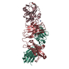 6p8nC 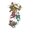 6mftS S: Starting model for refinement C: citing same article ( |
|---|---|
| Similar structure data |


-Links
 PDBj
PDBj


- Assembly
| Deposited unit | 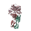
| ||||||||||||
|---|---|---|---|---|---|---|---|---|---|---|---|---|---|
| 1 |
| ||||||||||||
| Unit cell |
| ||||||||||||
| Components on special symmetry positions |
|
-Components
-Protein , 1 types, 1 molecules C
| #1: Protein | Mass: 38694.062 Da / Num. of mol.: 1 Source method: isolated from a genetically manipulated source Source: (gene. exp.) Human immunodeficiency virus / Cell line (production host): HEK 293s GNTI(-/-) / Production host:  Homo sapiens (human) / References: UniProt: M4Q8P8*PLUS Homo sapiens (human) / References: UniProt: M4Q8P8*PLUS |
|---|
-Antibody , 2 types, 2 molecules HL
| #2: Antibody | Mass: 25374.393 Da / Num. of mol.: 1 Source method: isolated from a genetically manipulated source Source: (gene. exp.) Homo sapiens (human) |
|---|---|
| #3: Antibody | Mass: 23704.377 Da / Num. of mol.: 1 Source method: isolated from a genetically manipulated source Source: (gene. exp.) Homo sapiens (human) |
-Sugars , 2 types, 10 molecules 
| #4: Polysaccharide | alpha-D-mannopyranose-(1-3)-[alpha-D-mannopyranose-(1-6)]beta-D-mannopyranose-(1-4)-2-acetamido-2- ...alpha-D-mannopyranose-(1-3)-[alpha-D-mannopyranose-(1-6)]beta-D-mannopyranose-(1-4)-2-acetamido-2-deoxy-beta-D-glucopyranose-(1-4)-2-acetamido-2-deoxy-beta-D-glucopyranose Source method: isolated from a genetically manipulated source |
|---|---|
| #5: Sugar | ChemComp-NAG /  Type: D-saccharide, beta linking / Mass: 221.208 Da / Num. of mol.: 9 Type: D-saccharide, beta linking / Mass: 221.208 Da / Num. of mol.: 9Source method: isolated from a genetically manipulated source Formula: C8H15NO6 |
-Non-polymers , 6 types, 25 molecules 


| #6: Chemical | ChemComp-NH4 /  Mass: 18.038 Da / Num. of mol.: 4 / Source method: obtained synthetically / Formula: H4N Mass: 18.038 Da / Num. of mol.: 4 / Source method: obtained synthetically / Formula: H4N#7: Chemical |  Mass: 62.068 Da / Num. of mol.: 2 / Source method: obtained synthetically / Formula: C2H6O2 Mass: 62.068 Da / Num. of mol.: 2 / Source method: obtained synthetically / Formula: C2H6O2#8: Chemical |  Mass: 106.120 Da / Num. of mol.: 2 / Source method: obtained synthetically / Formula: C4H10O3 Mass: 106.120 Da / Num. of mol.: 2 / Source method: obtained synthetically / Formula: C4H10O3#9: Chemical | ChemComp-NA /  Mass: 22.990 Da / Num. of mol.: 4 / Source method: obtained synthetically / Formula: Na Mass: 22.990 Da / Num. of mol.: 4 / Source method: obtained synthetically / Formula: Na#10: Chemical | ChemComp-CL / |  Mass: 35.453 Da / Num. of mol.: 1 / Source method: obtained synthetically / Formula: Cl Mass: 35.453 Da / Num. of mol.: 1 / Source method: obtained synthetically / Formula: Cl#11: Water | ChemComp-HOH / | Mass: 18.015 Da / Num. of mol.: 12 / Source method: isolated from a natural source / Formula: H2O |
|---|
-Details
| Has ligand of interest | N |
|---|---|
| Has protein modification | Y |
-Experimental details
-Experiment
| Experiment | Method: X-RAY DIFFRACTION / Number of used crystals: 1 |
|---|
- Sample preparation
| Crystal | Density Matthews: 4.01 Å3/Da / Density % sol: 69.34 % |
|---|---|
| Crystal grow | Temperature: 293 K / Method: vapor diffusion, sitting drop / pH: 5.5 / Details: 0.67% PEG 4000, 0.67M Ammonium Citrate pH 5.5 / Temp details: Room Temperature |
-Data collection
| Diffraction | Mean temperature: 100 K / Serial crystal experiment: N |
|---|---|
| Diffraction source | Source: SYNCHROTRON / Site: ALS / Beamline: 5.0.2 / Wavelength: 1 Å |
| Detector | Type: DECTRIS PILATUS3 6M / Detector: PIXEL / Date: Oct 4, 2018 |
| Radiation | Protocol: SINGLE WAVELENGTH / Monochromatic (M) / Laue (L): M / Scattering type: x-ray |
| Radiation wavelength | Wavelength: 1 Å / Relative weight: 1 |
| Reflection | Resolution: 3.59→50 Å / Num. obs: 11547 / % possible obs: 82.5 % / Redundancy: 2.8 % / CC1/2: 0.99 / Net I/σ(I): 7.75 |
| Reflection shell | Resolution: 3.59→3.65 Å / Redundancy: 2.4 % / Mean I/σ(I) obs: 1.075 / Num. unique obs: 468 / CC1/2: 0.801 / % possible all: 75.8 |
- Processing
| Software |
| |||||||||||||||||||||||||||||||||||
|---|---|---|---|---|---|---|---|---|---|---|---|---|---|---|---|---|---|---|---|---|---|---|---|---|---|---|---|---|---|---|---|---|---|---|---|---|
| Refinement | Method to determine structure: MOLECULAR REPLACEMENT Starting model: 6MFT Resolution: 3.594→50 Å / SU ML: 0.43 / Cross valid method: FREE R-VALUE / σ(F): 1.34 / Phase error: 27.38
| |||||||||||||||||||||||||||||||||||
| Solvent computation | Shrinkage radii: 0.9 Å / VDW probe radii: 1.11 Å | |||||||||||||||||||||||||||||||||||
| Refinement step | Cycle: LAST / Resolution: 3.594→50 Å
| |||||||||||||||||||||||||||||||||||
| Refine LS restraints |
| |||||||||||||||||||||||||||||||||||
| LS refinement shell |
|
