 Movie
Movie Controller

[English] 日本語
 Yorodumi
- PDB-2b4c: Crystal structure of HIV-1 JR-FL gp120 core protein containing th... -
+ Open data

- Basic information
| Entry | Database: PDB / ID: 2b4c | |||||||||
|---|---|---|---|---|---|---|---|---|---|---|
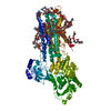
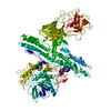
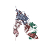
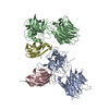
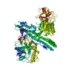
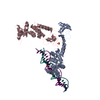
| Title | Crystal structure of HIV-1 JR-FL gp120 core protein containing the third variable region (V3) complexed with CD4 and the X5 antibody | |||||||||
 Components Components |
| |||||||||
 Keywords Keywords | Viral protein/Immune system / HIV-1 / gp120 / JRFL / V3 / X5 / CD4 induced antibody / Viral protein-Immune system COMPLEX | |||||||||
| Function / homology |  Function and homology information Function and homology informationhelper T cell enhancement of adaptive immune response / interleukin-16 binding / interleukin-16 receptor activity / maintenance of protein location in cell / cellular response to ionomycin / response to methamphetamine hydrochloride / T cell selection / MHC class II protein binding / interleukin-15-mediated signaling pathway / cellular response to granulocyte macrophage colony-stimulating factor stimulus ...helper T cell enhancement of adaptive immune response / interleukin-16 binding / interleukin-16 receptor activity / maintenance of protein location in cell / cellular response to ionomycin / response to methamphetamine hydrochloride / T cell selection / MHC class II protein binding / interleukin-15-mediated signaling pathway / cellular response to granulocyte macrophage colony-stimulating factor stimulus / positive regulation of monocyte differentiation / Alpha-defensins / Nef Mediated CD4 Down-regulation / regulation of T cell activation / response to vitamin D / Other interleukin signaling / extracellular matrix structural constituent / T cell receptor complex / enzyme-linked receptor protein signaling pathway / Translocation of ZAP-70 to Immunological synapse / Phosphorylation of CD3 and TCR zeta chains / Generation of second messenger molecules / macrophage differentiation / immunoglobulin binding / T cell differentiation / Co-inhibition by PD-1 / positive regulation of calcium ion transport into cytosol / Binding and entry of HIV virion / positive regulation of interleukin-2 production / coreceptor activity / symbiont-mediated perturbation of host defense response / positive regulation of plasma membrane raft polarization / positive regulation of receptor clustering / positive regulation of T cell proliferation / positive regulation of calcium-mediated signaling / cell surface receptor protein tyrosine kinase signaling pathway / protein tyrosine kinase binding / host cell endosome membrane / clathrin-coated endocytic vesicle membrane / calcium-mediated signaling / Vpu mediated degradation of CD4 / MHC class II protein complex binding / response to estradiol / transmembrane signaling receptor activity / Downstream TCR signaling / Cargo recognition for clathrin-mediated endocytosis / T cell receptor signaling pathway / Clathrin-mediated endocytosis / virus receptor activity / signaling receptor activity / clathrin-dependent endocytosis of virus by host cell / defense response to Gram-negative bacterium / adaptive immune response / response to ethanol / early endosome / cell surface receptor signaling pathway / cell adhesion / immune response / viral protein processing / membrane raft / endoplasmic reticulum lumen / fusion of virus membrane with host plasma membrane / external side of plasma membrane / fusion of virus membrane with host endosome membrane / viral envelope / symbiont entry into host cell / lipid binding / protein kinase binding / endoplasmic reticulum membrane / virion attachment to host cell / host cell plasma membrane / virion membrane / structural molecule activity / enzyme binding / signal transduction / protein homodimerization activity / zinc ion binding / membrane / identical protein binding / plasma membrane Similarity search - Function | |||||||||
| Biological species |   Human immunodeficiency virus 1 Human immunodeficiency virus 1 Homo sapiens (human) Homo sapiens (human) | |||||||||
| Method |  X-RAY DIFFRACTION / SYNCHROTRON / MOLECULAR REPLACEMENT / Resolution: 3.3 Å X-RAY DIFFRACTION / SYNCHROTRON / MOLECULAR REPLACEMENT / Resolution: 3.3 Å | |||||||||
 Authors Authors | Huang, C. / Tang, M. / Zhang, M.Y. / Majeed, S. / Montabana, E. / Stanfield, R.L. / Dimitrov, D.S. / Korber, B. / Sodroski, J. / Wilson, I.A. ...Huang, C. / Tang, M. / Zhang, M.Y. / Majeed, S. / Montabana, E. / Stanfield, R.L. / Dimitrov, D.S. / Korber, B. / Sodroski, J. / Wilson, I.A. / Wyatt, R. / Kwong, P.D. | |||||||||
 Citation Citation | Journal: Science / Year: 2005 Title: Structure of a V3-containing HIV-1 gp120 core. Authors: Huang, C.C. / Tang, M. / Zhang, M.Y. / Majeed, S. / Montabana, E. / Stanfield, R.L. / Dimitrov, D.S. / Korber, B. / Sodroski, J. / Wilson, I.A. / Wyatt, R. / Kwong, P.D. | |||||||||
| History |
|
- Structure visualization
| Structure viewer | Molecule: MolmilJmol/JSmol |
|---|
- Downloads & links
-Download
| PDBx/mmCIF format | 2b4c.cif.gz | 205.5 KB | Display | PDBx/mmCIF format |
|---|---|---|---|---|
| PDB format | pdb2b4c.ent.gz | 160.1 KB | Display | PDB format |
| PDBx/mmJSON format | 2b4c.json.gz | Tree view | PDBx/mmJSON format | |
| Others |  Other downloads Other downloads |
-Validation report
| Arichive directory | https://data.pdbj.org/pub/pdb/validation_reports/b4/2b4cftp://data.pdbj.org/pub/pdb/validation_reports/b4/2b4c | HTTPS FTP |
|---|
-Related structure data


-Links
 PDBj
PDBj


- Assembly
| Deposited unit | 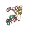
| |||||||||
|---|---|---|---|---|---|---|---|---|---|---|
| 1 |
| |||||||||
| 2 | 
| |||||||||
| Unit cell |
| |||||||||
| Components on special symmetry positions |
|
-Components
-Protein , 2 types, 2 molecules GC
| #1: Protein | Mass: 38452.652 Da / Num. of mol.: 1 / Fragment: CORE with V3 / Mutation: N301Q, T389A Source method: isolated from a genetically manipulated source Source: (gene. exp.) Human immunodeficiency virus 1 / Genus: Lentivirus / Production host:  |
|---|---|
| #2: Protein | Mass: 20129.896 Da / Num. of mol.: 1 / Fragment: D1D2, N-TERMINAL TWO DOMAIN FRAGMENT Source method: isolated from a genetically manipulated source Source: (gene. exp.) Homo sapiens (human) / Gene: CD4 / Production host:   Cricetulus griseus (Chinese hamster) / References: UniProt: P01730 Cricetulus griseus (Chinese hamster) / References: UniProt: P01730 |
-Antibody , 2 types, 2 molecules LH
| #3: Antibody | Mass: 23267.781 Da / Num. of mol.: 1 Source method: isolated from a genetically manipulated source Source: (gene. exp.) Homo sapiens (human) / Production host:  |
|---|---|
| #4: Antibody | Mass: 24990.732 Da / Num. of mol.: 1 Source method: isolated from a genetically manipulated source Source: (gene. exp.) Homo sapiens (human) / Production host: |
-Sugars , 3 types, 7 molecules 

| #5: Polysaccharide | alpha-L-fucopyranose-(1-6)-2-acetamido-2-deoxy-beta-D-glucopyranose Source method: isolated from a genetically manipulated source | ||
|---|---|---|---|
| #6: Sugar |  Type: D-saccharide, beta linking / Mass: 221.208 Da / Num. of mol.: 3 / Source method: obtained synthetically / Formula: C8H15NO6 Type: D-saccharide, beta linking / Mass: 221.208 Da / Num. of mol.: 3 / Source method: obtained synthetically / Formula: C8H15NO6#8: Sugar |  Type: D-saccharide / Mass: 152.146 Da / Num. of mol.: 3 / Source method: obtained synthetically / Formula: C5H12O5 Type: D-saccharide / Mass: 152.146 Da / Num. of mol.: 3 / Source method: obtained synthetically / Formula: C5H12O5 |
-Non-polymers , 1 types, 5 molecules 
| #7: Chemical | ChemComp-SO4 /  Mass: 96.063 Da / Num. of mol.: 5 / Source method: obtained synthetically / Formula: SO4 Mass: 96.063 Da / Num. of mol.: 5 / Source method: obtained synthetically / Formula: SO4 |
|---|
-Details
| Has protein modification | Y |
|---|
-Experimental details
-Experiment
| Experiment | Method: X-RAY DIFFRACTION / Number of used crystals: 1 |
|---|
- Sample preparation
| Crystal | Density Matthews: 3.28 Å3/Da / Density % sol: 62 % |
|---|---|
| Crystal grow | Temperature: 293 K / pH: 5.5 Details: ammonium sulfate, VAPOR DIFFUSION, HANGING DROP, temperature 293K, pH 5.50 |
-Data collection
| Diffraction | Mean temperature: 100 K |
|---|---|
| Diffraction source | Source: SYNCHROTRON / Site: APS  / Beamline: 22-ID / Wavelength: 1 / Beamline: 22-ID / Wavelength: 1 |
| Detector | Type: MARRESEARCH / Detector: CCD / Date: Mar 12, 2004 |
| Radiation | Monochromator: SI (220) / Protocol: SINGLE WAVELENGTH / Monochromatic (M) / Laue (L): M / Scattering type: x-ray |
| Radiation wavelength | Wavelength: 1 Å / Relative weight: 1 |
| Reflection | Resolution: 3.3→20 Å / Num. obs: 19372 / % possible obs: 86.6 % / Observed criterion σ(I): -3 / Redundancy: 9.6 % / Rmerge(I) obs: 0.082 / Rsym value: 0.082 / Net I/σ(I): 26.2 |
| Reflection shell | Resolution: 3.3→3.42 Å / Redundancy: 3.1 % / Mean I/σ(I) obs: 1.3 / Rsym value: 0.505 / % possible all: 20.9 |
- Processing
| Software |
| ||||||||||||||||||||||||||||||||||||||||||||||||||||||||||||
|---|---|---|---|---|---|---|---|---|---|---|---|---|---|---|---|---|---|---|---|---|---|---|---|---|---|---|---|---|---|---|---|---|---|---|---|---|---|---|---|---|---|---|---|---|---|---|---|---|---|---|---|---|---|---|---|---|---|---|---|---|---|
| Refinement | Method to determine structure: MOLECULAR REPLACEMENT Starting model: PDB ENTRY 1RHH AND 1RZK Resolution: 3.3→20 Å / Rfactor Rfree error: 0.008 / Data cutoff high absF: 8776198.43 / Data cutoff low absF: 0 / Cross valid method: THROUGHOUT / σ(F): 0 / Stereochemistry target values: ENGH & HUBER
| ||||||||||||||||||||||||||||||||||||||||||||||||||||||||||||
| Displacement parameters | Biso mean: 126.2 Å2
| ||||||||||||||||||||||||||||||||||||||||||||||||||||||||||||
| Refine analyze |
| ||||||||||||||||||||||||||||||||||||||||||||||||||||||||||||
| Refinement step | Cycle: LAST / Resolution: 3.3→20 Å
| ||||||||||||||||||||||||||||||||||||||||||||||||||||||||||||
| Refine LS restraints |
| ||||||||||||||||||||||||||||||||||||||||||||||||||||||||||||
| LS refinement shell | Resolution: 3.3→3.51 Å / Rfactor Rfree error: 0.043 / Total num. of bins used: 6
|
