ムービー
ムービー コントローラー


+ データを開く

- 基本情報
基本情報
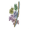
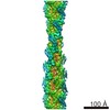
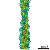
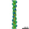
| 登録情報 | データベース: PDB / ID: 3jbi | ||||||
|---|---|---|---|---|---|---|---|
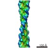
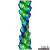
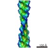
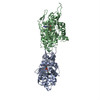
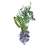
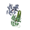
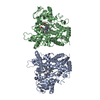
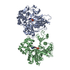
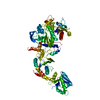
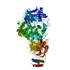
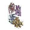
| タイトル | MDFF model of the vinculin tail domain bound to F-actin | ||||||
 要素 要素 |
| ||||||
 キーワード キーワード |  STRUCTURAL PROTEIN (タンパク質) / cytoskeleton (細胞骨格) / adhesion (接着) STRUCTURAL PROTEIN (タンパク質) / cytoskeleton (細胞骨格) / adhesion (接着) | ||||||
| 機能・相同性 |  機能・相同性情報 機能・相同性情報muscle tendon junction / Platelet degranulation / Smooth Muscle Contraction / regulation of protein localization to adherens junction / podosome ring / outer dense plaque of desmosome / inner dense plaque of desmosome / terminal web / epithelial cell-cell adhesion / 接着結合 ...muscle tendon junction / Platelet degranulation / Smooth Muscle Contraction / regulation of protein localization to adherens junction / podosome ring / outer dense plaque of desmosome / inner dense plaque of desmosome / terminal web / epithelial cell-cell adhesion / 接着結合 / dystroglycan binding / muscle alpha-actinin binding / MAP2K and MAPK activation / alpha-catenin binding / vinculin binding / fascia adherens / cell-cell contact zone / apical junction assembly / costamere / regulation of establishment of endothelial barrier / adherens junction assembly / axon extension / protein localization to cell surface / lamellipodium assembly / cytoskeletal motor activator activity / regulation of focal adhesion assembly / tropomyosin binding / myosin heavy chain binding / mesenchyme migration / troponin I binding / actin filament bundle / filamentous actin / alpha-actinin binding / skeletal muscle thin filament assembly / actin filament bundle assembly / striated muscle thin filament / 刷子縁 / skeletal muscle myofibril / actin monomer binding / skeletal muscle fiber development / stress fiber / titin binding / regulation of cell migration / actin filament polymerization / filopodium / cell projection / Neutrophil degranulation / マイクロフィラメント / morphogenesis of an epithelium / 接着結合 / 加水分解酵素; 酸無水物に作用; 酸無水物に作用・細胞または細胞小器官の運動に関与 / 筋鞘 / neuromuscular junction / beta-catenin binding / Z disc / calcium-dependent protein binding / actin filament binding / cell-cell junction / マイクロフィラメント / lamellipodium / cell body / scaffold protein binding / ミトコンドリア内膜 / 細胞骨格 / hydrolase activity / 細胞接着 / cadherin binding / protein domain specific binding / focal adhesion / ubiquitin protein ligase binding / calcium ion binding / positive regulation of gene expression / structural molecule activity / magnesium ion binding / protein homodimerization activity / protein-containing complex / ATP binding / identical protein binding / 細胞膜 / 細胞質類似検索 - 分子機能 | ||||||
| 生物種 |  Gallus gallus (ニワトリ) Gallus gallus (ニワトリ) Oryctolagus cuniculus (ウサギ) Oryctolagus cuniculus (ウサギ) | ||||||
| 手法 | 電子顕微鏡法 / らせん対称体再構成法 / クライオ電子顕微鏡法 / 解像度: 8.5 Å | ||||||
 データ登録者 データ登録者 | Kim, L.Y. / Thompson, P.M. / Lee, H.T. / Pershad, M. / Campbell, S.L. / Alushin, G.M. | ||||||
 引用 引用 | ジャーナル: J Mol Biol / 年: 2016 タイトル: The Structural Basis of Actin Organization by Vinculin and Metavinculin. 著者: Laura Y Kim / Peter M Thompson / Hyunna T Lee / Mihir Pershad / Sharon L Campbell / Gregory M Alushin /  要旨: Vinculin is an essential adhesion protein that links membrane-bound integrin and cadherin receptors through their intracellular binding partners to filamentous actin, facilitating mechanotransduction. ...Vinculin is an essential adhesion protein that links membrane-bound integrin and cadherin receptors through their intracellular binding partners to filamentous actin, facilitating mechanotransduction. Here we present an 8.5-Å-resolution cryo-electron microscopy reconstruction and pseudo-atomic model of the vinculin tail (Vt) domain bound to F-actin. Upon actin engagement, the N-terminal "strap" and helix 1 are displaced from the Vt helical bundle to mediate actin bundling. We find that an analogous conformational change also occurs in the H1' helix of the tail domain of metavinculin (MVt) upon actin binding, a muscle-specific splice isoform that suppresses actin bundling by Vt. These data support a model in which metavinculin tunes the actin bundling activity of vinculin in a tissue-specific manner, providing a mechanistic framework for understanding metavinculin mutations associated with hereditary cardiomyopathies. | ||||||
| 履歴 |
|
- 構造の表示
構造の表示
| ムービー |
ムービービューア |
|---|---|
| 構造ビューア | 分子: MolmilJmol/JSmol |
- ダウンロードとリンク
-ダウンロード
| PDBx/mmCIF形式 | 3jbi.cif.gz | 163 KB | 表示 | PDBx/mmCIF形式 |
|---|---|---|---|---|
| PDB形式 | pdb3jbi.ent.gz | 122.1 KB | 表示 | PDB形式 |
| PDBx/mmJSON形式 | 3jbi.json.gz | ツリー表示 | PDBx/mmJSON形式 | |
| その他 |  その他のダウンロード その他のダウンロード |
-検証レポート
| アーカイブディレクトリ | https://data.pdbj.org/pub/pdb/validation_reports/jb/3jbiftp://data.pdbj.org/pub/pdb/validation_reports/jb/3jbi | HTTPS FTP |
|---|
-関連構造データ


-リンク
 PDBj
PDBj


- 集合体
集合体
| 登録構造単位 | 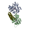
|
|---|---|
| 1 |
|
| 対称性 | らせん対称: (回転対称性: 1 / Dyad axis: no / N subunits divisor: 1 / Num. of operations: 1 / Rise per n subunits: 27.8 Å / Rotation per n subunits: -166.82 °) |
-要素
| #1: タンパク質 | / Alpha-actin-1 分子量: 41862.613 Da / 分子数: 2 / 由来タイプ: 天然 / 由来: (天然) Oryctolagus cuniculus (ウサギ) / 組織: skeletal muscle骨格筋 / 参照: UniProt: P68135#2: タンパク質 | | ビンキュリン / Vt分子量: 28241.018 Da / 分子数: 1 / Fragment: tail domain (UNP residues 879-1130) / 由来タイプ: 組換発現 / 由来: (組換発現) Gallus gallus (ニワトリ) / 遺伝子: VCL, VINC1 / Organelle: focal adhesion / 発現宿主:  Escherichia coli BL21(DE3) (大腸菌) / 参照: UniProt: P12003 Escherichia coli BL21(DE3) (大腸菌) / 参照: UniProt: P12003#3: 化合物 |   分子量: 24.305 Da / 分子数: 2 / 由来タイプ: 合成 / 式: Mg 分子量: 24.305 Da / 分子数: 2 / 由来タイプ: 合成 / 式: Mg#4: 化合物 | アデノシン二リン酸  分子量: 427.201 Da / 分子数: 2 / 由来タイプ: 合成 / 式: C10H15N5O10P2 / コメント: ADP, エネルギー貯蔵分子*YM 分子量: 427.201 Da / 分子数: 2 / 由来タイプ: 合成 / 式: C10H15N5O10P2 / コメント: ADP, エネルギー貯蔵分子*YM |
|---|
-実験情報
-実験
| 実験 | 手法: 電子顕微鏡法 |
|---|---|
| EM実験 | 試料の集合状態: FILAMENT / 3次元再構成法: らせん対称体再構成法 |
- 試料調製
試料調製
| 構成要素 |
| ||||||||||||||||
|---|---|---|---|---|---|---|---|---|---|---|---|---|---|---|---|---|---|
| 緩衝液 | 名称: 50 mM KCl, 1 mM MgCl2, 1 mM EGTA, 10 mM imidazole / pH: 7 / 詳細: 50 mM KCl, 1 mM MgCl2, 1 mM EGTA, 10 mM imidazole | ||||||||||||||||
| 試料 | 濃度: 0.0125 mg/ml / 包埋: NO / シャドウイング: NO / 染色: NO / 凍結: YES | ||||||||||||||||
| 試料支持 | 詳細: 200 mesh 1.2 / 1.3 C-flat | ||||||||||||||||
| 急速凍結 | 装置: LEICA EM GP / 凍結剤: ETHANE / 湿度: 90 % 詳細: 3 microliters of 0.3 micromolar actin was applied to the grid and incubated for 60 seconds at 25 degrees C. 3 microliters of 10 micromolar Vt was then applied and incubated for 60 seconds. 3 ...詳細: 3 microliters of 0.3 micromolar actin was applied to the grid and incubated for 60 seconds at 25 degrees C. 3 microliters of 10 micromolar Vt was then applied and incubated for 60 seconds. 3 microliters of solution was removed, then an additional 3 microliters of Vt applied. After 60 seconds, 3 microliters of solution was removed, then the grid was blotted for 2 seconds before plunging into liquid ethane (LEICA EM GP). 手法: 3 microliters of 0.3 micromolar actin was applied to the grid and incubated for 60 seconds at 25 degrees C. 3 microliters of 10 micromolar Vt was then applied and incubated for 60 seconds. 3 ...手法: 3 microliters of 0.3 micromolar actin was applied to the grid and incubated for 60 seconds at 25 degrees C. 3 microliters of 10 micromolar Vt was then applied and incubated for 60 seconds. 3 microliters of solution was removed, then an additional 3 microliters of Vt applied. After 60 seconds, 3 microliters of solution was removed, then the grid was blotted for 2 seconds before plunging. |
- 電子顕微鏡撮影
| 実験機器 |  モデル: Tecnai F20 / 画像提供: FEI Company |
|---|---|
| 顕微鏡 | モデル: FEI TECNAI F20 / 日付: 2014年5月22日 |
| 電子銃 | 電子線源: FIELD EMISSION GUN / 加速電圧: 120 kV / 照射モード: FLOOD BEAM |
| 電子レンズ | モード: BRIGHT FIELDBright-field microscopy / 倍率(公称値): 100000 X / 倍率(補正後): 137615 X / 最大 デフォーカス(公称値): 3000 nm / 最小 デフォーカス(公称値): 1500 nm / Cs: 2 mm 非点収差 : Objective lens astigmatism was corrected at 100,000 times magnification. |
| 試料ホルダ | 試料ホルダーモデル: GATAN LIQUID NITROGEN |
| 撮影 | 電子線照射量: 25 e/Å2 フィルム・検出器のモデル: GATAN ULTRASCAN 4000 (4k x 4k) |
| 画像スキャン | デジタル画像の数: 1384 |
- 解析
解析
| EMソフトウェア |
| ||||||||||||||||||||||||||||
|---|---|---|---|---|---|---|---|---|---|---|---|---|---|---|---|---|---|---|---|---|---|---|---|---|---|---|---|---|---|
| CTF補正 | 詳細: FREALIGN (per segment) | ||||||||||||||||||||||||||||
| らせん対称 | 回転角度/サブユニット: 166.82 ° / 軸方向距離/サブユニット: 27.8 Å / らせん対称軸の対称性: C1 | ||||||||||||||||||||||||||||
| 3次元再構成 | 手法: IHRSR / 解像度: 8.5 Å / 解像度の算出法: FSC 0.143 CUT-OFF / ピクセルサイズ(公称値): 2.18 Å / ピクセルサイズ(実測値): 2.18 Å / 倍率補正: catalase crystal 詳細: (Helical Details: Single model IHRSR was performed with EMAN2 / SPARX and final reconstruction with FREALIGN (fixed helical parameters).) 対称性のタイプ: HELICAL | ||||||||||||||||||||||||||||
| 原子モデル構築 |
| ||||||||||||||||||||||||||||
| 原子モデル構築 | PDB chain-ID: A / Source name: PDB / タイプ: experimental model
| ||||||||||||||||||||||||||||
| 精密化ステップ | サイクル: LAST
|