 Movie
Movie Controller

[English] 日本語
 Yorodumi
- EMDB-6450: Unsharpened cryo-EM reconstruction of the Vt-GFP-actin interface ... -
+ Open data

- Basic information
| Entry | Database: EMDB / ID: EMD-6450 | |||||||||
|---|---|---|---|---|---|---|---|---|---|---|
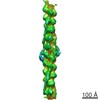
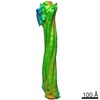
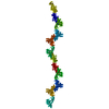
| Title | Unsharpened cryo-EM reconstruction of the Vt-GFP-actin interface for difference map calculation | |||||||||
 Map data Map data | Unsharpened reconstruction of C-terminal GFP fusion to the vinculin tail domain (Vt-GFP) bound to actin for difference map calculation | |||||||||
 Sample Sample |
| |||||||||
 Keywords Keywords | actin / vinculin / cell migration / adhesion / mechanosensation / cytoskeleton | |||||||||
| Function / homology |  Function and homology information Function and homology informationmuscle tendon junction / Platelet degranulation / Smooth Muscle Contraction / regulation of protein localization to adherens junction / outer dense plaque of desmosome / inner dense plaque of desmosome / podosome ring / terminal web / epithelial cell-cell adhesion / zonula adherens ...muscle tendon junction / Platelet degranulation / Smooth Muscle Contraction / regulation of protein localization to adherens junction / outer dense plaque of desmosome / inner dense plaque of desmosome / podosome ring / terminal web / epithelial cell-cell adhesion / zonula adherens / Regulation of CDH1 Function / fascia adherens / MAP2K and MAPK activation / dystroglycan binding / alpha-catenin binding / muscle alpha-actinin binding / vinculin binding / cell-cell contact zone / apical junction assembly / costamere / regulation of establishment of endothelial barrier / adherens junction assembly / protein localization to cell surface / lamellipodium assembly / regulation of focal adhesion assembly / cytoskeletal motor activator activity / myosin heavy chain binding / tropomyosin binding / actin filament bundle / troponin I binding / filamentous actin / mesenchyme migration / skeletal muscle myofibril / alpha-actinin binding / brush border / actin filament bundle assembly / striated muscle thin filament / skeletal muscle thin filament assembly / actin monomer binding / skeletal muscle fiber development / stress fiber / titin binding / actin filament polymerization / regulation of cell migration / Neutrophil degranulation / morphogenesis of an epithelium / filopodium / actin filament / adherens junction / neuromuscular junction / sarcolemma / beta-catenin binding / Hydrolases; Acting on acid anhydrides; Acting on acid anhydrides to facilitate cellular and subcellular movement / Z disc / calcium-dependent protein binding / cell-cell junction / actin filament binding / lamellipodium / actin cytoskeleton / cell body / scaffold protein binding / cytoskeleton / cell adhesion / mitochondrial inner membrane / cadherin binding / protein domain specific binding / focal adhesion / hydrolase activity / calcium ion binding / positive regulation of gene expression / ubiquitin protein ligase binding / perinuclear region of cytoplasm / structural molecule activity / magnesium ion binding / protein homodimerization activity / protein-containing complex / ATP binding / identical protein binding / plasma membrane / cytoplasm Similarity search - Function | |||||||||
| Biological species |   | |||||||||
| Method | helical reconstruction / cryo EM / Resolution: 19.2 Å | |||||||||
 Authors Authors | Kim LY / Thompson PM / Lee HT / Pershad M / Campbell SL / Alushin GM | |||||||||
 Citation Citation | Journal: J Mol Biol / Year: 2016 Title: The Structural Basis of Actin Organization by Vinculin and Metavinculin. Authors: Laura Y Kim / Peter M Thompson / Hyunna T Lee / Mihir Pershad / Sharon L Campbell / Gregory M Alushin /  Abstract: Vinculin is an essential adhesion protein that links membrane-bound integrin and cadherin receptors through their intracellular binding partners to filamentous actin, facilitating mechanotransduction. ...Vinculin is an essential adhesion protein that links membrane-bound integrin and cadherin receptors through their intracellular binding partners to filamentous actin, facilitating mechanotransduction. Here we present an 8.5-Å-resolution cryo-electron microscopy reconstruction and pseudo-atomic model of the vinculin tail (Vt) domain bound to F-actin. Upon actin engagement, the N-terminal "strap" and helix 1 are displaced from the Vt helical bundle to mediate actin bundling. We find that an analogous conformational change also occurs in the H1' helix of the tail domain of metavinculin (MVt) upon actin binding, a muscle-specific splice isoform that suppresses actin bundling by Vt. These data support a model in which metavinculin tunes the actin bundling activity of vinculin in a tissue-specific manner, providing a mechanistic framework for understanding metavinculin mutations associated with hereditary cardiomyopathies. | |||||||||
| History |
|
- Structure visualization
| Movie |
Movie viewer |
|---|---|
| Structure viewer | EM map: SurfViewMolmilJmol/JSmol |
| Supplemental images |

- Downloads & links
-EMDB archive
| Map data | emd_6450.map.gz | 4 MB | EMDB map data format | |
|---|---|---|---|---|
| Header (meta data) | emd-6450-v30.xmlemd-6450.xml | 11.6 KB 11.6 KB | Display Display | EMDB header |
| Images | 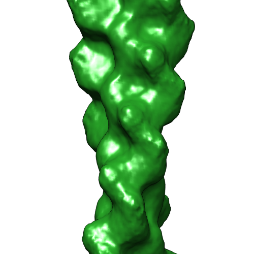 emd_6450.png emd_6450.png | 126.1 KB | ||
| Archive directory |  http://ftp.pdbj.org/pub/emdb/structures/EMD-6450ftp://ftp.pdbj.org/pub/emdb/structures/EMD-6450 http://ftp.pdbj.org/pub/emdb/structures/EMD-6450ftp://ftp.pdbj.org/pub/emdb/structures/EMD-6450 | HTTPS FTP |
-Related structure data
| Related structure data |  6446C  6447C  6448C 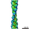 6449C 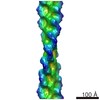 6451C 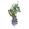 3jbiC 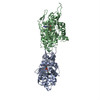 3jbjC 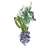 3jbkC C: citing same article ( |
|---|---|
| Similar structure data |
-Links
| EMDB pages | EMDB (EBI/PDBe) / EMDataResource |
|---|---|
| Related items in Molecule of the Month |


-Map
| File | Download / File: emd_6450.map.gz / Format: CCP4 / Size: 62.5 MB / Type: IMAGE STORED AS FLOATING POINT NUMBER (4 BYTES) | ||||||||||||||||||||||||||||||||||||||||||||||||||||||||||||
|---|---|---|---|---|---|---|---|---|---|---|---|---|---|---|---|---|---|---|---|---|---|---|---|---|---|---|---|---|---|---|---|---|---|---|---|---|---|---|---|---|---|---|---|---|---|---|---|---|---|---|---|---|---|---|---|---|---|---|---|---|---|
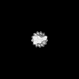
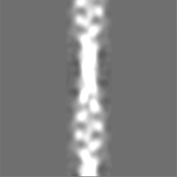
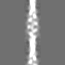
| Annotation | Unsharpened reconstruction of C-terminal GFP fusion to the vinculin tail domain (Vt-GFP) bound to actin for difference map calculation | ||||||||||||||||||||||||||||||||||||||||||||||||||||||||||||
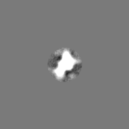
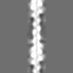
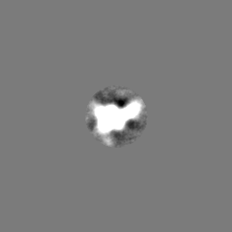
| Projections & slices | Image control
Images are generated by Spider. | ||||||||||||||||||||||||||||||||||||||||||||||||||||||||||||
| Voxel size | X=Y=Z: 2.18 Å | ||||||||||||||||||||||||||||||||||||||||||||||||||||||||||||
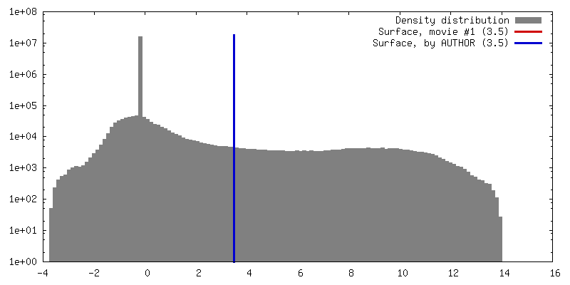
| Density |
| ||||||||||||||||||||||||||||||||||||||||||||||||||||||||||||
| Symmetry | Space group: 1 | ||||||||||||||||||||||||||||||||||||||||||||||||||||||||||||
| Details | EMDB XML:
CCP4 map header:
| ||||||||||||||||||||||||||||||||||||||||||||||||||||||||||||
 Z (Sec.)
 Y (Row.)
 X (Col.)


-Supplemental data
- Sample components
-Entire : Vt-GFP bound to F-actin
| Entire | Name: Vt-GFP bound to F-actin |
|---|---|
| Components |
|
-Supramolecule #1000: Vt-GFP bound to F-actin
| Supramolecule | Name: Vt-GFP bound to F-actin / type: sample / ID: 1000 Details: Helical filament with one vinculin tail domain bound per actin protomer Oligomeric state: One vinculin tail domain per actin protomer Number unique components: 2 |
|---|
-Macromolecule #1: skeletal muscle actin
| Macromolecule | Name: skeletal muscle actin / type: protein_or_peptide / ID: 1 / Name.synonym: actin / Oligomeric state: helical filament / Recombinant expression: No / Database: NCBI |
|---|---|
| Source (natural) | Organism: |
| Molecular weight | Theoretical: 41.8 KDa |
| Sequence | UniProtKB: Actin, alpha skeletal muscle |
-Macromolecule #2: Vinculin tail domain, residues 879-1061
| Macromolecule | Name: Vinculin tail domain, residues 879-1061 / type: protein_or_peptide / ID: 2 / Name.synonym: Vt-GFP / Details: C-terminal GFP fusion / Oligomeric state: helical / Recombinant expression: Yes |
|---|---|
| Source (natural) | Organism: |
| Molecular weight | Theoretical: 55.1 KDa |
| Recombinant expression | Organism:  |
| Sequence | UniProtKB: Vinculin |
-Experimental details
-Structure determination
| Method | cryo EM |
|---|---|
 Processing Processing | helical reconstruction |
| Aggregation state | filament |
-Sample preparation
| Concentration | 0.0125 mg/mL |
|---|---|
| Buffer | pH: 7 / Details: 50 mM KCl, 1 mM MgCl2, 1 mM EGTA, 10 mM imidazole |
| Grid | Details: 200 mesh 1.2 / 1.3 C-flat |
| Vitrification | Cryogen name: ETHANE / Chamber humidity: 90 % / Instrument: LEICA EM GP Method: 3 microliters of 0.3 micromolar actin was applied to the grid and incubated for 60 seconds at 25 degrees C. 3 microliters of 10 micromolar Vt-GFP was then applied and incubated for 60 seconds. ...Method: 3 microliters of 0.3 micromolar actin was applied to the grid and incubated for 60 seconds at 25 degrees C. 3 microliters of 10 micromolar Vt-GFP was then applied and incubated for 60 seconds. 3 microliters of solution was removed, then an additional 3 microliters of Vt-GFP applied. After 60 seconds, 3 microliters of solution was removed, then the grid was blotted for 2 seconds before plunging. |
- Electron microscopy
| Microscope | FEI TECNAI 20 |
|---|---|
| Alignment procedure | Legacy - Astigmatism: Objective lens astigmatism was corrected at 100,000 times magnification. |
| Date | May 12, 2014 |
| Image recording | Category: CCD / Film or detector model: GATAN ULTRASCAN 4000 (4k x 4k) / Number real images: 260 / Average electron dose: 25 e/Å2 |
| Electron beam | Acceleration voltage: 120 kV / Electron source:  FIELD EMISSION GUN FIELD EMISSION GUN |
| Electron optics | Calibrated magnification: 137615 / Illumination mode: FLOOD BEAM / Imaging mode: BRIGHT FIELD / Cs: 2.0 mm / Nominal defocus max: 3.0 µm / Nominal defocus min: 1.5 µm / Nominal magnification: 100000 |
| Sample stage | Specimen holder model: GATAN LIQUID NITROGEN |
-Image processing
| Details | Multi-model IHRSR was performed using EMAN2 / SPARX to select for bound segments, followed by single-model IHRSR with EMAN2 / SPARX and final reconstruction with FREALIGN (fixed helical parameters). |
|---|---|
| Final reconstruction | Applied symmetry - Helical parameters - Δz: 27.87 Å Applied symmetry - Helical parameters - Δ&Phi: 166.60 ° Applied symmetry - Helical parameters - Axial symmetry: C1 (asymmetric) Algorithm: OTHER / Resolution.type: BY AUTHOR / Resolution: 19.2 Å / Resolution method: OTHER / Software - Name: CTFFIND3, EMAN2/SPARX, FREALIGN |
| CTF correction | Details: FREALIGN (per segment) |
