 Movie
Movie Controller

[English] 日本語
 Yorodumi
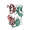
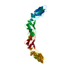
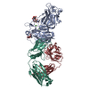
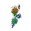
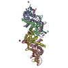
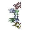
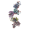
- PDB-1uj3: Crystal structure of a humanized Fab fragment of anti-tissue-fact... -
+ Open data

- Basic information
| Entry | Database: PDB / ID: 1uj3 | ||||||
|---|---|---|---|---|---|---|---|
| Title | Crystal structure of a humanized Fab fragment of anti-tissue-factor antibody in complex with tissue factor | ||||||
 Components Components |
| ||||||
 Keywords Keywords |  IMMUNE SYSTEM/BLOOD CLOTTING / tissue factor / antigen / antibody / Fab / IMMUNE SYSTEM-BLOOD CLOTTING COMPLEX IMMUNE SYSTEM/BLOOD CLOTTING / tissue factor / antigen / antibody / Fab / IMMUNE SYSTEM-BLOOD CLOTTING COMPLEX | ||||||
| Function / homology |  Function and homology information Function and homology informationactivation of plasma proteins involved in acute inflammatory response / activation of blood coagulation via clotting cascade / serine-type peptidase complex / positive regulation of platelet-derived growth factor receptor signaling pathway / NGF-stimulated transcription / cytokine receptor activity / positive regulation of positive chemotaxis / Extrinsic Pathway of Fibrin Clot Formation / positive regulation of TOR signaling / positive regulation of endothelial cell proliferation ...activation of plasma proteins involved in acute inflammatory response / activation of blood coagulation via clotting cascade / serine-type peptidase complex / positive regulation of platelet-derived growth factor receptor signaling pathway / NGF-stimulated transcription / cytokine receptor activity / positive regulation of positive chemotaxis / Extrinsic Pathway of Fibrin Clot Formation / positive regulation of TOR signaling / positive regulation of endothelial cell proliferation / positive regulation of interleukin-8 production / phospholipid binding / protein processing / cytokine-mediated signaling pathway / activation of cysteine-type endopeptidase activity involved in apoptotic process / positive regulation of angiogenesis / blood coagulation / collagen-containing extracellular matrix / protease binding / positive regulation of cell migration / external side of plasma membrane / positive regulation of gene expression / cell surface / extracellular space / membrane / plasma membraneSimilarity search - Function | ||||||
| Biological species |  Homo sapiens (human) Homo sapiens (human) | ||||||
| Method | X-RAY DIFFRACTION / SYNCHROTRON / MOLECULAR REPLACEMENT / Resolution: 2.1 Å | ||||||
 Authors Authors | Ohto, U. / Mizutani, R. / Nakamura, M. / Adachi, H. / Satow, Y. | ||||||
 Citation Citation | Journal: J.Synchrotron Radiat. / Year: 2004 Title: Crystal structure of a humanized Fab fragment of anti-tissue-factor antibody in complex with tissue factor. Authors: Ohto, U. / Mizutani, R. / Nakamura, M. / Adachi, H. / Satow, Y. | ||||||
| History |
|
- Structure visualization
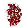
| Structure viewer | Molecule: MolmilJmol/JSmol |
|---|
- Downloads & links
-Download
| PDBx/mmCIF format | 1uj3.cif.gz | 147 KB | Display | PDBx/mmCIF format |
|---|---|---|---|---|
| PDB format | pdb1uj3.ent.gz | 112.8 KB | Display | PDB format |
| PDBx/mmJSON format | 1uj3.json.gz | Tree view | PDBx/mmJSON format | |
| Others |  Other downloads Other downloads |
-Validation report
| Arichive directory | https://data.pdbj.org/pub/pdb/validation_reports/uj/1uj3ftp://data.pdbj.org/pub/pdb/validation_reports/uj/1uj3 | HTTPS FTP |
|---|
-Related structure data
-Links
 PDBj
PDBj


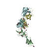
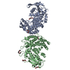
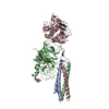
- Assembly
| Deposited unit | 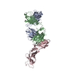
| ||||||||
|---|---|---|---|---|---|---|---|---|---|
| 1 |
| ||||||||
| Unit cell |
|
-Components
| #1: Antibody | Mass: 23475.979 Da / Num. of mol.: 1 / Fragment: anti-tissue-factor antibody hATR-5 Fab Source method: isolated from a genetically manipulated source Source: (gene. exp.) Homo sapiens (human) / Production host:  Escherichia coli (E. coli) Escherichia coli (E. coli) |
|---|---|
| #2: Antibody | Mass: 23255.990 Da / Num. of mol.: 1 / Fragment: anti-tissue-factor antibody hATR-5 Fab Source method: isolated from a genetically manipulated source Source: (gene. exp.) Homo sapiens (human) / Production host: Escherichia coli (E. coli) |
| #3: Protein | / TF / Coagulation factor III / Thromboplastin / CD142 antigen / blood coagulation factor Mass: 23302.949 Da / Num. of mol.: 1 / Fragment: RESIDUES 606-810 Source method: isolated from a genetically manipulated source Source: (gene. exp.) Homo sapiens (human) / Production host: Escherichia coli (E. coli) / References: UniProt: P13726 |
| #4: Water | ChemComp-HOH / Water Mass: 18.015 Da / Num. of mol.: 464 / Source method: isolated from a natural source / Formula: H2O Mass: 18.015 Da / Num. of mol.: 464 / Source method: isolated from a natural source / Formula: H2O |
-Experimental details
-Experiment
| Experiment | Method: X-RAY DIFFRACTION / Number of used crystals: 1 |
|---|
- Sample preparation
| Crystal | Density Matthews: 3.85 Å3/Da / Density % sol: 67.77 % |
|---|---|
| Crystal grow | Temperature: 277 K / Method: vapor diffusion, hanging drop / pH: 7.5 Details: 2-propanol, PEG4000, HEPES-Na, pH 7.5, VAPOR DIFFUSION, HANGING DROP, temperature 277K |
-Data collection
| Diffraction | Mean temperature: 100 K |
|---|---|
| Diffraction source | Source: SYNCHROTRON / Site: SPring-8  / Beamline: BL38B1 / Wavelength: 1 Å / Beamline: BL38B1 / Wavelength: 1 Å |
| Detector | Type: ADSC QUANTUM 4 / Detector: CCD / Date: Jul 5, 2002 |
| Radiation | Monochromator: SI 111 / Protocol: SINGLE WAVELENGTH / Monochromatic (M) / Laue (L): M / Scattering type: x-ray |
| Radiation wavelength | Wavelength: 1 Å / Relative weight: 1 |
| Reflection | Resolution: 2.1→37.1 Å / Num. all: 69013 / Num. obs: 64872 / % possible obs: 94 % / Observed criterion σ(F): 3 / Biso Wilson estimate: 19.2 Å2 / Rmerge(I) obs: 0.087 |
| Reflection shell | Resolution: 2.1→2.2 Å / % possible all: 81 |
- Processing
| Software |
| ||||||||||||||||||||||||||||||||||||||||||||||||||||||||||||||||||||||||||||||||
|---|---|---|---|---|---|---|---|---|---|---|---|---|---|---|---|---|---|---|---|---|---|---|---|---|---|---|---|---|---|---|---|---|---|---|---|---|---|---|---|---|---|---|---|---|---|---|---|---|---|---|---|---|---|---|---|---|---|---|---|---|---|---|---|---|---|---|---|---|---|---|---|---|---|---|---|---|---|---|---|---|---|
| Refinement | Method to determine structure: MOLECULAR REPLACEMENT Starting model: PDB ENTRIES 2HFT AND 1JPT Resolution: 2.1→37.01 Å / Rfactor Rfree error: 0.003 / Data cutoff high absF: 1947766.14 / Data cutoff low absF: 0 / Isotropic thermal model: RESTRAINED / Cross valid method: THROUGHOUT / σ(F): 3 / Stereochemistry target values: Engh & Huber
| ||||||||||||||||||||||||||||||||||||||||||||||||||||||||||||||||||||||||||||||||
| Solvent computation | Solvent model: FLAT MODEL / Bsol: 44.9528 Å2 / ksol: 0.339345 e/Å3 | ||||||||||||||||||||||||||||||||||||||||||||||||||||||||||||||||||||||||||||||||
| Displacement parameters | Biso mean: 32.3 Å2
| ||||||||||||||||||||||||||||||||||||||||||||||||||||||||||||||||||||||||||||||||
| Refine analyze |
| ||||||||||||||||||||||||||||||||||||||||||||||||||||||||||||||||||||||||||||||||
| Refinement step | Cycle: LAST / Resolution: 2.1→37.01 Å
| ||||||||||||||||||||||||||||||||||||||||||||||||||||||||||||||||||||||||||||||||
| Refine LS restraints |
| ||||||||||||||||||||||||||||||||||||||||||||||||||||||||||||||||||||||||||||||||
| LS refinement shell | Resolution: 2.1→2.23 Å / Rfactor Rfree error: 0.008 / Total num. of bins used: 6
| ||||||||||||||||||||||||||||||||||||||||||||||||||||||||||||||||||||||||||||||||
| Xplor file |
|
