 Movie
Movie Controller


+ Open data

- Basic information
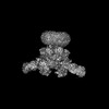
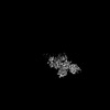
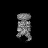
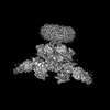
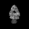
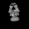
| Entry | Database: PDB / ID: 8vut | ||||||
|---|---|---|---|---|---|---|---|
| Title | Human GluN1-2A with IgG 008-218 | ||||||
 Components Components |
| ||||||
 Keywords Keywords | MEMBRANE PROTEIN/IMMUNE SYSTEM / Channel / heterotetramer / receptor / antibody / MEMBRANE PROTEIN-IMMUNE SYSTEM complex | ||||||
| Function / homology |  Function and homology information Function and homology informationexcitatory chemical synaptic transmission / directional locomotion / Synaptic adhesion-like molecules / serotonin metabolic process / protein localization to postsynaptic membrane / propylene metabolic process / response to glycine / sleep / activation of cysteine-type endopeptidase activity / Assembly and cell surface presentation of NMDA receptors ...excitatory chemical synaptic transmission / directional locomotion / Synaptic adhesion-like molecules / serotonin metabolic process / protein localization to postsynaptic membrane / propylene metabolic process / response to glycine / sleep / activation of cysteine-type endopeptidase activity / Assembly and cell surface presentation of NMDA receptors / regulation of monoatomic cation transmembrane transport / glutamate receptor signaling pathway / NMDA glutamate receptor activity / Neurexins and neuroligins / NMDA selective glutamate receptor complex / calcium ion transmembrane import into cytosol / glutamate binding / protein heterotetramerization / positive regulation of reactive oxygen species biosynthetic process / positive regulation of calcium ion transport into cytosol / glycine binding / dopamine metabolic process / Negative regulation of NMDA receptor-mediated neuronal transmission / Unblocking of NMDA receptors, glutamate binding and activation / startle response / monoatomic cation transmembrane transport / regulation of neuronal synaptic plasticity / monoatomic cation transport / Long-term potentiation / positive regulation of excitatory postsynaptic potential / ligand-gated monoatomic ion channel activity / excitatory synapse / calcium ion homeostasis / MECP2 regulates neuronal receptors and channels / synaptic cleft / glutamate-gated calcium ion channel activity / sensory perception of pain / EPHB-mediated forward signaling / Ras activation upon Ca2+ influx through NMDA receptor / response to amphetamine / ionotropic glutamate receptor signaling pathway / neurogenesis / positive regulation of synaptic transmission, glutamatergic / regulation of membrane potential / excitatory postsynaptic potential / transmitter-gated monoatomic ion channel activity involved in regulation of postsynaptic membrane potential / synaptic membrane / synaptic transmission, glutamatergic / long-term synaptic potentiation / postsynaptic density membrane / negative regulation of protein catabolic process / visual learning / cytoplasmic vesicle membrane / protein catabolic process / brain development / regulation of synaptic plasticity / terminal bouton / memory / response to wounding / synaptic vesicle / signaling receptor activity / presynaptic membrane / amyloid-beta binding / RAF/MAP kinase cascade / chemical synaptic transmission / postsynaptic membrane / response to ethanol / dendritic spine / postsynaptic density / learning or memory / calmodulin binding / neuron projection / response to xenobiotic stimulus / positive regulation of apoptotic process / glutamatergic synapse / calcium ion binding / dendrite / synapse / endoplasmic reticulum membrane / protein-containing complex binding / cell surface / positive regulation of transcription by RNA polymerase II / zinc ion binding / plasma membrane / cytoplasm Similarity search - Function | ||||||
| Biological species |  Homo sapiens (human) Homo sapiens (human) | ||||||
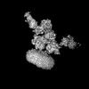
| Method | ELECTRON MICROSCOPY / single particle reconstruction / cryo EM / Resolution: 3.7 Å | ||||||
 Authors Authors | Michalski, K. / Furukawa, H. | ||||||
| Funding support |  United States, 1items United States, 1items
| ||||||
 Citation Citation | Journal: Nat Struct Mol Biol / Year: 2024 Title: Structural and functional mechanisms of anti-NMDAR autoimmune encephalitis. Authors: Kevin Michalski / Taha Abdulla / Sam Kleeman / Lars Schmidl / Ricardo Gómez / Noriko Simorowski / Francesca Vallese / Harald Prüss / Manfred Heckmann / Christian Geis / Hiro Furukawa /  Abstract: Autoantibodies against neuronal membrane proteins can manifest in autoimmune encephalitis, inducing seizures, cognitive dysfunction and psychosis. Anti-N-methyl-D-aspartate receptor (NMDAR) ...Autoantibodies against neuronal membrane proteins can manifest in autoimmune encephalitis, inducing seizures, cognitive dysfunction and psychosis. Anti-N-methyl-D-aspartate receptor (NMDAR) encephalitis is the most dominant autoimmune encephalitis; however, insights into how autoantibodies recognize and alter receptor functions remain limited. Here we determined structures of human and rat NMDARs bound to three distinct patient-derived antibodies using single-particle electron cryo-microscopy. These antibodies bind different regions within the amino-terminal domain of the GluN1 subunit. Through electrophysiology, we show that all three autoantibodies acutely and directly reduced NMDAR channel functions in primary neurons. Antibodies show different stoichiometry of binding and antibody-receptor complex formation, which in one antibody, 003-102, also results in reduced synaptic localization of NMDARs. These studies demonstrate mechanisms of diverse epitope recognition and direct channel regulation of anti-NMDAR autoantibodies underlying autoimmune encephalitis. | ||||||
| History |
|
- Structure visualization
| Structure viewer | Molecule: MolmilJmol/JSmol |
|---|
- Downloads & links
-Download
| PDBx/mmCIF format | 8vut.cif.gz | 659.5 KB | Display | PDBx/mmCIF format |
|---|---|---|---|---|
| PDB format | pdb8vut.ent.gz | 522.5 KB | Display | PDB format |
| PDBx/mmJSON format | 8vut.json.gz | Tree view | PDBx/mmJSON format | |
| Others |  Other downloads Other downloads |
-Validation report
| Summary document | 8vut_validation.pdf.gz | 1.4 MB | Display | wwPDB validaton report |
|---|---|---|---|---|
| Full document | 8vut_full_validation.pdf.gz | 1.4 MB | Display | |
| Data in XML | 8vut_validation.xml.gz | 107 KB | Display | |
| Data in CIF | 8vut_validation.cif.gz | 164.8 KB | Display | |
| Arichive directory | https://data.pdbj.org/pub/pdb/validation_reports/vu/8vutftp://data.pdbj.org/pub/pdb/validation_reports/vu/8vut | HTTPS FTP |
-Related structure data
| Related structure data | 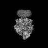 43539MC 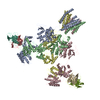 8vuhC 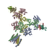 8vujC 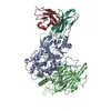 8vulC 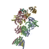 8vunC 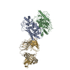 8vuqC 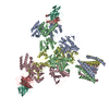 8vurC 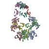 8vusC 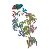 8vuuC 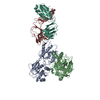 8vuvC  8vuyC 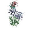 8vvhC M: map data used to model this data C: citing same article ( |
|---|---|
| Similar structure data |

-Links
 PDBj
PDBj


- Assembly
| Deposited unit | 
|
|---|---|
| 1 |
|
-Components
| #1: Protein | Mass: 91923.094 Da / Num. of mol.: 2 Source method: isolated from a genetically manipulated source Source: (gene. exp.) Homo sapiens (human) / Gene: GRIN1, NMDAR1 / Production host:   Spodoptera frugiperda (fall armyworm) / References: UniProt: Q05586 Spodoptera frugiperda (fall armyworm) / References: UniProt: Q05586#2: Protein | Mass: 90704.898 Da / Num. of mol.: 2 Source method: isolated from a genetically manipulated source Source: (gene. exp.) Homo sapiens (human) / Gene: GRIN2A, NMDAR2A / Production host: Spodoptera frugiperda (fall armyworm) / References: UniProt: Q12879#3: Antibody | Mass: 23367.164 Da / Num. of mol.: 2 Source method: isolated from a genetically manipulated source Source: (gene. exp.) Homo sapiens (human) / Production host: Homo sapiens (human)#4: Protein | Mass: 22380.652 Da / Num. of mol.: 2 Source method: isolated from a genetically manipulated source Source: (gene. exp.) Homo sapiens (human) / Production host: Homo sapiens (human) |
|---|
-Experimental details
-Experiment
| Experiment | Method: ELECTRON MICROSCOPY |
|---|---|
| EM experiment | Aggregation state: PARTICLE / 3D reconstruction method: single particle reconstruction |
- Sample preparation
| Component | Name: Human GluN1-2A with IgG 008-218 / Type: COMPLEX / Entity ID: all / Source: RECOMBINANT |
|---|---|
| Molecular weight | Experimental value: NO |
| Source (natural) | Organism: Homo sapiens (human) |
| Source (recombinant) | Organism: Spodoptera frugiperda (fall armyworm) |
| Buffer solution | pH: 7.5 |
| Specimen | Embedding applied: NO / Shadowing applied: NO / Staining applied: NO / Vitrification applied: YES |
| Vitrification | Cryogen name: ETHANE |
- Electron microscopy imaging
| Experimental equipment |  Model: Titan Krios / Image courtesy: FEI Company |
|---|---|
| Microscopy | Model: FEI TITAN KRIOS |
| Electron gun | Electron source:  FIELD EMISSION GUN / Accelerating voltage: 300 kV / Illumination mode: OTHER FIELD EMISSION GUN / Accelerating voltage: 300 kV / Illumination mode: OTHER |
| Electron lens | Mode: OTHER / Nominal defocus max: 2200 nm / Nominal defocus min: 800 nm / Cs: 2.7 mm |
| Image recording | Electron dose: 60 e/Å2 / Film or detector model: GATAN K3 (6k x 4k) |
- Processing
| EM software | Name: cryoSPARC / Category: 3D reconstruction | ||||||||||||||||||||||||
|---|---|---|---|---|---|---|---|---|---|---|---|---|---|---|---|---|---|---|---|---|---|---|---|---|---|
| CTF correction | Type: NONE | ||||||||||||||||||||||||
| 3D reconstruction | Resolution: 3.7 Å / Resolution method: FSC 0.143 CUT-OFF / Num. of particles: 287872 / Symmetry type: POINT | ||||||||||||||||||||||||
| Refine LS restraints |
|
