 Movie
Movie Controller

[English] 日本語
 Yorodumi
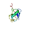
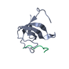
- PDB-7wrp: Crystal Structure of pks13-ACP domain from Corynebacterium diphtheriae -
+ Open data

- Basic information
| Entry | Database: PDB / ID: 7wrp | ||||||
|---|---|---|---|---|---|---|---|
| Title | Crystal Structure of pks13-ACP domain from Corynebacterium diphtheriae | ||||||
 Components Components | Polyketide synthase involved in mycolic acid biosynthesis | ||||||
 Keywords Keywords | RECOMBINATION / acyl-carrier protein | ||||||
| Function / homology | 4'-PHOSPHOPANTETHEINE / Polyketide synthase involved in mycolic acid biosynthesis Function and homology information Function and homology information | ||||||
| Biological species |  Corynebacterium diphtheriae (bacteria) Corynebacterium diphtheriae (bacteria) | ||||||
| Method |  X-RAY DIFFRACTION / SYNCHROTRON / MOLECULAR REPLACEMENT / Resolution: 1.794 Å X-RAY DIFFRACTION / SYNCHROTRON / MOLECULAR REPLACEMENT / Resolution: 1.794 Å | ||||||
 Authors Authors | Liu, X. | ||||||
| Funding support | 1items
| ||||||
 Citation Citation | Journal: Biochem.Biophys.Res.Commun. / Year: 2022 Title: Crystal structures of FadD32 and pks13-ACP domain from Corynebacterium diphtheriae. Authors: Chen, R. / Yuan, J. / Shi, X. / Tang, W. / Liu, X. | ||||||
| History |
|
- Structure visualization
| Structure viewer | Molecule: MolmilJmol/JSmol |
|---|
- Downloads & links
-Download
| PDBx/mmCIF format | 7wrp.cif.gz | 32.3 KB | Display | PDBx/mmCIF format |
|---|---|---|---|---|
| PDB format | pdb7wrp.ent.gz | 19.6 KB | Display | PDB format |
| PDBx/mmJSON format | 7wrp.json.gz | Tree view | PDBx/mmJSON format | |
| Others |  Other downloads Other downloads |
-Validation report
| Arichive directory | https://data.pdbj.org/pub/pdb/validation_reports/wr/7wrpftp://data.pdbj.org/pub/pdb/validation_reports/wr/7wrp | HTTPS FTP |
|---|
-Related structure data
| Related structure data |  6c4qS S: Starting model for refinement |
|---|---|
| Similar structure data |


-Links
 PDBj
PDBj- Assembly
| Deposited unit | 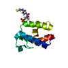
| ||||||||
|---|---|---|---|---|---|---|---|---|---|
| 1 |
| ||||||||
| Unit cell |
|
-Components
| #1: Protein | Mass: 9349.608 Da / Num. of mol.: 1 / Source method: obtained synthetically / Source: (synth.) Corynebacterium diphtheriae (bacteria) / References: UniProt: A0A806GVW0 |
|---|---|
| #2: Chemical | ChemComp-PNS / 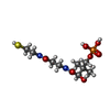  Mass: 358.348 Da / Num. of mol.: 1 / Source method: obtained synthetically / Formula: C11H23N2O7PS / Feature type: SUBJECT OF INVESTIGATION Mass: 358.348 Da / Num. of mol.: 1 / Source method: obtained synthetically / Formula: C11H23N2O7PS / Feature type: SUBJECT OF INVESTIGATION |
| #3: Water | ChemComp-HOH /  Mass: 18.015 Da / Num. of mol.: 90 / Source method: isolated from a natural source / Formula: H2O Mass: 18.015 Da / Num. of mol.: 90 / Source method: isolated from a natural source / Formula: H2O |
| Has ligand of interest | Y |
-Experimental details
-Experiment
| Experiment | Method: X-RAY DIFFRACTION / Number of used crystals: 1 |
|---|
- Sample preparation
| Crystal | Density Matthews: 3.57 Å3/Da / Density % sol: 65.59 % / Mosaicity: 0.83 ° |
|---|---|
| Crystal grow | Temperature: 293 K / Method: evaporation / pH: 8 Details: 0.2 M (NH4)2SO4, 0.1 M NaAc??3H2O pH 4.6, 25% w/v PEG 4,000, VAPOR DIFFUSION, Sitting DROP, temperature 293K |
-Data collection
| Diffraction | Mean temperature: 100 K / Serial crystal experiment: N | ||||||||||||||||||||||||||||||||||||||||||||||||||||||||||||||||||||||||||||||||||||||||||||||||||||||||||||||||||||||||||||||||||||||||||||||||||||||||||||||||||||||||
|---|---|---|---|---|---|---|---|---|---|---|---|---|---|---|---|---|---|---|---|---|---|---|---|---|---|---|---|---|---|---|---|---|---|---|---|---|---|---|---|---|---|---|---|---|---|---|---|---|---|---|---|---|---|---|---|---|---|---|---|---|---|---|---|---|---|---|---|---|---|---|---|---|---|---|---|---|---|---|---|---|---|---|---|---|---|---|---|---|---|---|---|---|---|---|---|---|---|---|---|---|---|---|---|---|---|---|---|---|---|---|---|---|---|---|---|---|---|---|---|---|---|---|---|---|---|---|---|---|---|---|---|---|---|---|---|---|---|---|---|---|---|---|---|---|---|---|---|---|---|---|---|---|---|---|---|---|---|---|---|---|---|---|---|---|---|---|---|---|---|
| Diffraction source | Source: SYNCHROTRON / Site: SSRF  / Beamline: BL19U1 / Wavelength: 0.9784 Å / Beamline: BL19U1 / Wavelength: 0.9784 Å | ||||||||||||||||||||||||||||||||||||||||||||||||||||||||||||||||||||||||||||||||||||||||||||||||||||||||||||||||||||||||||||||||||||||||||||||||||||||||||||||||||||||||
| Detector | Type: DECTRIS PILATUS 6M / Detector: PIXEL / Date: May 14, 2021 | ||||||||||||||||||||||||||||||||||||||||||||||||||||||||||||||||||||||||||||||||||||||||||||||||||||||||||||||||||||||||||||||||||||||||||||||||||||||||||||||||||||||||
| Radiation | Protocol: SINGLE WAVELENGTH / Monochromatic (M) / Laue (L): M / Scattering type: x-ray | ||||||||||||||||||||||||||||||||||||||||||||||||||||||||||||||||||||||||||||||||||||||||||||||||||||||||||||||||||||||||||||||||||||||||||||||||||||||||||||||||||||||||
| Radiation wavelength | Wavelength: 0.9784 Å / Relative weight: 1 | ||||||||||||||||||||||||||||||||||||||||||||||||||||||||||||||||||||||||||||||||||||||||||||||||||||||||||||||||||||||||||||||||||||||||||||||||||||||||||||||||||||||||
| Reflection | Resolution: 1.794→50 Å / Num. obs: 13451 / % possible obs: 100 % / Redundancy: 22.2 % / Rmerge(I) obs: 0.121 / Rpim(I) all: 0.028 / Rrim(I) all: 0.125 / Χ2: 1.209 / Net I/σ(I): 7.4 | ||||||||||||||||||||||||||||||||||||||||||||||||||||||||||||||||||||||||||||||||||||||||||||||||||||||||||||||||||||||||||||||||||||||||||||||||||||||||||||||||||||||||
| Reflection shell | Diffraction-ID: 1 / % possible all: 100
|
- Processing
| Software |
| |||||||||||||||||||||||||
|---|---|---|---|---|---|---|---|---|---|---|---|---|---|---|---|---|---|---|---|---|---|---|---|---|---|---|
| Refinement | Method to determine structure: MOLECULAR REPLACEMENT Starting model: 6c4q Resolution: 1.794→34.568 Å / SU ML: 0.18 / Cross valid method: THROUGHOUT / σ(F): 1.36 / Phase error: 27 / Stereochemistry target values: ML
| |||||||||||||||||||||||||
| Solvent computation | Shrinkage radii: 0.9 Å / VDW probe radii: 1.11 Å / Solvent model: FLAT BULK SOLVENT MODEL | |||||||||||||||||||||||||
| Displacement parameters | Biso max: 123.98 Å2 / Biso mean: 37.7649 Å2 / Biso min: 20.32 Å2 | |||||||||||||||||||||||||
| Refinement step | Cycle: final / Resolution: 1.794→34.568 Å
| |||||||||||||||||||||||||
| LS refinement shell | Refine-ID: X-RAY DIFFRACTION / Rfactor Rfree error: 0 / % reflection obs: 100 %
|
