 Movie
Movie Controller


+ Open data

- Basic information
| Entry | Database: PDB / ID: 1n5z | ||||||
|---|---|---|---|---|---|---|---|
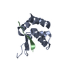
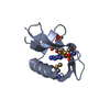
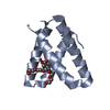
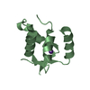
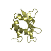
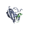
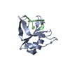
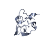
| Title | Complex structure of Pex13p SH3 domain with a peptide of Pex14p | ||||||
 Components Components |
| ||||||
 Keywords Keywords | PROTEIN TRANSPORT / SH3 domain / PxxP motif | ||||||
| Function / homology |  Function and homology information Function and homology informationprotein import into peroxisome matrix, translocation / peroxisomal importomer complex / Class I peroxisomal membrane protein import / Peroxisomal protein import / protein import into peroxisome matrix, docking / : / peroxisomal membrane / transmembrane protein transporter activity / protein-macromolecule adaptor activity / signaling receptor binding Similarity search - Function | ||||||
| Biological species |  | ||||||
| Method |  X-RAY DIFFRACTION / SYNCHROTRON / MOLECULAR REPLACEMENT / Resolution: 2.7 Å X-RAY DIFFRACTION / SYNCHROTRON / MOLECULAR REPLACEMENT / Resolution: 2.7 Å | ||||||
 Authors Authors | Douangamath, A. / Filipp, F.V. / Klein, A.T.J. / Barnett, P. / Zou, P. / Voorn-Brouwer, T. / Vega, M.C. / Mayans, O.M. / Sattler, M. / Distel, B. / Wilmanns, M. | ||||||
 Citation Citation | Journal: MOL.CELL / Year: 2002 Title: Topography for Independent Binding of alpha-Helical and PPII-Helical Ligands to a Peroxisomal SH3 Domain Authors: Douangamath, A. / Filipp, F.V. / Klein, A.T.J. / Barnett, P. / Zou, P. / Voorn-Brouwer, T. / Vega, M.C. / Mayans, O.M. / Sattler, M. / Distel, B. / Wilmanns, M. | ||||||
| History |
|
- Structure visualization
| Structure viewer | Molecule: MolmilJmol/JSmol |
|---|
- Downloads & links
-Download
| PDBx/mmCIF format | 1n5z.cif.gz | 46.2 KB | Display | PDBx/mmCIF format |
|---|---|---|---|---|
| PDB format | pdb1n5z.ent.gz | 34 KB | Display | PDB format |
| PDBx/mmJSON format | 1n5z.json.gz | Tree view | PDBx/mmJSON format | |
| Others |  Other downloads Other downloads |
-Validation report
| Arichive directory | https://data.pdbj.org/pub/pdb/validation_reports/n5/1n5zftp://data.pdbj.org/pub/pdb/validation_reports/n5/1n5z | HTTPS FTP |
|---|
-Related structure data


-Links
 PDBj
PDBj

- Assembly
| Deposited unit | 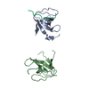
| ||||||||
|---|---|---|---|---|---|---|---|---|---|
| 1 | 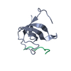
| ||||||||
| 2 | 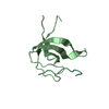
| ||||||||
| 3 | 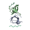
| ||||||||
| 4 | 
| ||||||||
| Unit cell |
|
-Components
| #1: Protein | Mass: 10782.284 Da / Num. of mol.: 2 / Fragment: SH3 domain Source method: isolated from a genetically manipulated source Source: (gene. exp.) Plasmid: pMALc2 / Species (production host): Escherichia coli / Production host:  #2: Protein/peptide | Mass: 1695.914 Da / Num. of mol.: 2 / Source method: obtained synthetically Details: Chemically synthesized. This sequence occurs naturally in Saccharomyces cerevisiae. References: UniProt: P53112 #3: Water | ChemComp-HOH / |  Mass: 18.015 Da / Num. of mol.: 18 / Source method: isolated from a natural source / Formula: H2O Mass: 18.015 Da / Num. of mol.: 18 / Source method: isolated from a natural source / Formula: H2O |
|---|
-Experimental details
-Experiment
| Experiment | Method: X-RAY DIFFRACTION / Number of used crystals: 1 |
|---|
- Sample preparation
| Crystal | Density Matthews: 2.83 Å3/Da / Density % sol: 56.2 % | ||||||||||||||||||||||||||||||||||||
|---|---|---|---|---|---|---|---|---|---|---|---|---|---|---|---|---|---|---|---|---|---|---|---|---|---|---|---|---|---|---|---|---|---|---|---|---|---|
| Crystal grow | Temperature: 298 K / Method: vapor diffusion, sitting drop / pH: 7.3 Details: Ammonium sulfate, lithium sulfate, tris, pH 7.3, VAPOR DIFFUSION, SITTING DROP, temperature 298K | ||||||||||||||||||||||||||||||||||||
| Crystal grow | *PLUS Temperature: 22 ℃ / Method: vapor diffusion | ||||||||||||||||||||||||||||||||||||
| Components of the solutions | *PLUS
|
-Data collection
| Diffraction | Mean temperature: 200 K |
|---|---|
| Diffraction source | Source: SYNCHROTRON / Site: EMBL/DESY, HAMBURG  / Beamline: X31 / Wavelength: 1.1 Å / Beamline: X31 / Wavelength: 1.1 Å |
| Detector | Type: MARRESEARCH / Detector: IMAGE PLATE / Date: Dec 10, 2001 |
| Radiation | Monochromator: double crystal monochromator / Protocol: SINGLE WAVELENGTH / Monochromatic (M) / Laue (L): M / Scattering type: x-ray |
| Radiation wavelength | Wavelength: 1.1 Å / Relative weight: 1 |
| Reflection | Resolution: 2.7→25 Å / Num. all: 6298 / Num. obs: 5907 / % possible obs: 93.8 % / Observed criterion σ(F): 0 / Observed criterion σ(I): 0 / Redundancy: 4.3 % / Biso Wilson estimate: 65 Å2 / Rsym value: 0.0065 / Net I/σ(I): 21.1 |
| Reflection shell | Highest resolution: 2.7 Å / Redundancy: 4.3 % / Mean I/σ(I) obs: 2.9 / Rsym value: 0.033 / % possible all: 97.4 |
| Reflection | *PLUS Highest resolution: 2.7 Å / Lowest resolution: 25 Å / Rmerge(I) obs: 0.065 |
| Reflection shell | *PLUS % possible obs: 97.4 % / Rmerge(I) obs: 0.33 |
- Processing
| Software |
| |||||||||||||||||||||||||||||||||||||||||||||||||||||||||||||||||||||||||||||||||||||||||||||||||||||||||||||||||||||||||||||
|---|---|---|---|---|---|---|---|---|---|---|---|---|---|---|---|---|---|---|---|---|---|---|---|---|---|---|---|---|---|---|---|---|---|---|---|---|---|---|---|---|---|---|---|---|---|---|---|---|---|---|---|---|---|---|---|---|---|---|---|---|---|---|---|---|---|---|---|---|---|---|---|---|---|---|---|---|---|---|---|---|---|---|---|---|---|---|---|---|---|---|---|---|---|---|---|---|---|---|---|---|---|---|---|---|---|---|---|---|---|---|---|---|---|---|---|---|---|---|---|---|---|---|---|---|---|---|
| Refinement | Method to determine structure: MOLECULAR REPLACEMENT / Resolution: 2.7→25 Å / Cor.coef. Fo:Fc: 0.901 / Cor.coef. Fo:Fc free: 0.894 / SU B: 13.558 / SU ML: 0.278 / Cross valid method: THROUGHOUT / σ(F): 0 / ESU R: 1.36 / ESU R Free: 0.371 / Stereochemistry target values: MAXIMUM LIKELIHOOD
| |||||||||||||||||||||||||||||||||||||||||||||||||||||||||||||||||||||||||||||||||||||||||||||||||||||||||||||||||||||||||||||
| Solvent computation | Shrinkage radii: 0.8 Å / Solvent model: BABINET MODEL WITH MASK | |||||||||||||||||||||||||||||||||||||||||||||||||||||||||||||||||||||||||||||||||||||||||||||||||||||||||||||||||||||||||||||
| Displacement parameters | Biso mean: 7.974 Å2
| |||||||||||||||||||||||||||||||||||||||||||||||||||||||||||||||||||||||||||||||||||||||||||||||||||||||||||||||||||||||||||||
| Refinement step | Cycle: LAST / Resolution: 2.7→25 Å
| |||||||||||||||||||||||||||||||||||||||||||||||||||||||||||||||||||||||||||||||||||||||||||||||||||||||||||||||||||||||||||||
| Refine LS restraints |
| |||||||||||||||||||||||||||||||||||||||||||||||||||||||||||||||||||||||||||||||||||||||||||||||||||||||||||||||||||||||||||||
| LS refinement shell | Resolution: 2.7→2.769 Å / Total num. of bins used: 20 /
| |||||||||||||||||||||||||||||||||||||||||||||||||||||||||||||||||||||||||||||||||||||||||||||||||||||||||||||||||||||||||||||
| Refinement TLS params. | Method: refined / Refine-ID: X-RAY DIFFRACTION
| |||||||||||||||||||||||||||||||||||||||||||||||||||||||||||||||||||||||||||||||||||||||||||||||||||||||||||||||||||||||||||||
| Refinement TLS group |
| |||||||||||||||||||||||||||||||||||||||||||||||||||||||||||||||||||||||||||||||||||||||||||||||||||||||||||||||||||||||||||||
| Refinement | *PLUS % reflection Rfree: 5 % / Rfactor Rfree: 0.308 / Rfactor Rwork: 0.234 | |||||||||||||||||||||||||||||||||||||||||||||||||||||||||||||||||||||||||||||||||||||||||||||||||||||||||||||||||||||||||||||
| Solvent computation | *PLUS | |||||||||||||||||||||||||||||||||||||||||||||||||||||||||||||||||||||||||||||||||||||||||||||||||||||||||||||||||||||||||||||
| Displacement parameters | *PLUS | |||||||||||||||||||||||||||||||||||||||||||||||||||||||||||||||||||||||||||||||||||||||||||||||||||||||||||||||||||||||||||||
| Refine LS restraints | *PLUS
|
