 Movie
Movie Controller

[English] 日本語
 Yorodumi
- PDB-3nrl: Crystal Structure of protein RUMGNA_01417 from Ruminococcus gnavu... -
+ Open data

- Basic information
| Entry | Database: PDB / ID: 3nrl | ||||||
|---|---|---|---|---|---|---|---|
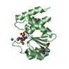
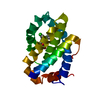
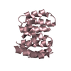
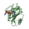
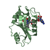
| Title | Crystal Structure of protein RUMGNA_01417 from Ruminococcus gnavus, Northeast Structural Genomics Consortium Target UgR76 | ||||||
 Components Components | uncharacterized protein RUMGNA_01417 | ||||||
 Keywords Keywords | structural genomics / unknown function / beta protein / PSI-2 / Protein Structure Initiative / Northeast Structural Genomics Consortium / NESG | ||||||
| Function / homology | Thrombin, subunit H - #390 / Domain of unknown function DUF5348 / Domain of unknown function (DUF5348) / Thrombin, subunit H / Beta Barrel / Mainly Beta / DUF5348 domain-containing protein Function and homology information Function and homology information | ||||||
| Biological species |  Ruminococcus gnavus (bacteria) Ruminococcus gnavus (bacteria) | ||||||
| Method |  X-RAY DIFFRACTION / SYNCHROTRON / SAD / Resolution: 1.9 Å X-RAY DIFFRACTION / SYNCHROTRON / SAD / Resolution: 1.9 Å | ||||||
 Authors Authors | Seetharaman, J. / Abashidze, M. / Sahdev, S. / Xiao, R. / Ciccosanti, C. / Lee, D. / Everett, J.K. / Nair, R. / Acton, T.B. / Rost, B. ...Seetharaman, J. / Abashidze, M. / Sahdev, S. / Xiao, R. / Ciccosanti, C. / Lee, D. / Everett, J.K. / Nair, R. / Acton, T.B. / Rost, B. / Montelione, G.T. / Tong, L. / Hunt, J.F. / Northeast Structural Genomics Consortium (NESG) | ||||||
 Citation Citation | Journal: To be Published Title: Northeast Structural Genomics Consortium Target UgR76 Authors: Seetharaman, J. / Abashidze, M. / Sahdev, S. / Xiao, R. / Ciccosanti, C. / Lee, D. / Everett, J.K. / Nair, R. / Acton, T.B. / Rost, B. / Montelione, G.T. / Tong, L. / Hunt, J.F. | ||||||
| History |
|
- Structure visualization
| Structure viewer | Molecule: MolmilJmol/JSmol |
|---|
- Downloads & links
-Download
| PDBx/mmCIF format | 3nrl.cif.gz | 43.2 KB | Display | PDBx/mmCIF format |
|---|---|---|---|---|
| PDB format | pdb3nrl.ent.gz | 30.4 KB | Display | PDB format |
| PDBx/mmJSON format | 3nrl.json.gz | Tree view | PDBx/mmJSON format | |
| Others |  Other downloads Other downloads |
-Validation report
| Arichive directory | https://data.pdbj.org/pub/pdb/validation_reports/nr/3nrlftp://data.pdbj.org/pub/pdb/validation_reports/nr/3nrl | HTTPS FTP |
|---|
-Related structure data
| Similar structure data | |
|---|---|
| Other databases |


-Links
 PDBj
PDBj- Assembly
| Deposited unit | 
| ||||||||
|---|---|---|---|---|---|---|---|---|---|
| 1 |
| ||||||||
| 2 | 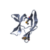
| ||||||||
| 3 | 
| ||||||||
| Unit cell |
| ||||||||
| Details | dimer |
-Components
| #1: Protein | Mass: 9817.574 Da / Num. of mol.: 2 Source method: isolated from a genetically manipulated source Source: (gene. exp.) Ruminococcus gnavus (bacteria) / Strain: ATCC 29149 / Gene: RUMGNA_01417 / Plasmid: pET 21-23C / Production host: #2: Chemical |   Mass: 96.063 Da / Num. of mol.: 2 / Source method: obtained synthetically / Formula: SO4 Mass: 96.063 Da / Num. of mol.: 2 / Source method: obtained synthetically / Formula: SO4#3: Water | ChemComp-HOH / |  Mass: 18.015 Da / Num. of mol.: 80 / Source method: isolated from a natural source / Formula: H2O Mass: 18.015 Da / Num. of mol.: 80 / Source method: isolated from a natural source / Formula: H2OHas protein modification | Y | |
|---|
-Experimental details
-Experiment
| Experiment | Method: X-RAY DIFFRACTION / Number of used crystals: 1 |
|---|
- Sample preparation
| Crystal | Density Matthews: 2.1 Å3/Da / Density % sol: 41.35 % |
|---|---|
| Crystal grow | Temperature: 291 K / Method: vapor diffusion, hanging drop / pH: 6.5 Details: Protein solution: 100mM NaCl, 5mM DTT, 0.02% NaN3, 10mM Tris-HCl (pH 7.5) . Reservoir solution: 0.1M Cacodylate Acid (pH 6.5), 18% PEG3350, and 0.2M ammonium sulfate., VAPOR DIFFUSION, ...Details: Protein solution: 100mM NaCl, 5mM DTT, 0.02% NaN3, 10mM Tris-HCl (pH 7.5) . Reservoir solution: 0.1M Cacodylate Acid (pH 6.5), 18% PEG3350, and 0.2M ammonium sulfate., VAPOR DIFFUSION, HANGING DROP, temperature 291K |
-Data collection
| Diffraction | Mean temperature: 100 K |
|---|---|
| Diffraction source | Source: SYNCHROTRON / Site: NSLS  / Beamline: X4C / Wavelength: 0.97915 Å / Beamline: X4C / Wavelength: 0.97915 Å |
| Detector | Type: MAR CCD 165 mm / Detector: CCD / Date: Jun 7, 2010 / Details: mirrors |
| Radiation | Monochromator: Si 111 CHANNEL / Protocol: SINGLE WAVELENGTH / Monochromatic (M) / Laue (L): M / Scattering type: x-ray |
| Radiation wavelength | Wavelength: 0.97915 Å / Relative weight: 1 |
| Reflection | Resolution: 1.9→50 Å / Num. all: 25141 / Num. obs: 24739 / % possible obs: 98.4 % / Observed criterion σ(F): 0 / Observed criterion σ(I): 0 / Redundancy: 4.3 % / Biso Wilson estimate: 10.8 Å2 / Rmerge(I) obs: 0.059 / Rsym value: 0.045 / Net I/σ(I): 29 |
| Reflection shell | Resolution: 1.9→1.93 Å / Redundancy: 3.7 % / Rmerge(I) obs: 0.365 / Mean I/σ(I) obs: 3.1 / Num. unique all: 1245 / Rsym value: 0.301 / % possible all: 91.2 |
- Processing
| Software |
| ||||||||||||||||||||||||||||||||
|---|---|---|---|---|---|---|---|---|---|---|---|---|---|---|---|---|---|---|---|---|---|---|---|---|---|---|---|---|---|---|---|---|---|
| Refinement | Method to determine structure: SAD / Resolution: 1.9→32.24 Å / Rfactor Rfree error: 0.006 / Occupancy max: 1 / Occupancy min: 1 / Data cutoff high absF: 171398 / Data cutoff low absF: 0 / Isotropic thermal model: RESTRAINED / Cross valid method: THROUGHOUT / σ(F): 2 / σ(I): 0 / Stereochemistry target values: Engh & Huber / Details: BULK SOLVENT MODEL USED
| ||||||||||||||||||||||||||||||||
| Solvent computation | Solvent model: FLAT MODEL / Bsol: 47.744 Å2 / ksol: 0.4 e/Å3 | ||||||||||||||||||||||||||||||||
| Displacement parameters | Biso max: 88.32 Å2 / Biso mean: 30.113 Å2 / Biso min: 9.58 Å2
| ||||||||||||||||||||||||||||||||
| Refine analyze |
| ||||||||||||||||||||||||||||||||
| Refinement step | Cycle: LAST / Resolution: 1.9→32.24 Å
| ||||||||||||||||||||||||||||||||
| Refine LS restraints |
| ||||||||||||||||||||||||||||||||
| LS refinement shell | Resolution: 1.9→2.02 Å / Rfactor Rfree error: 0.016 / Total num. of bins used: 6
| ||||||||||||||||||||||||||||||||
| Xplor file |
|
