 Movie
Movie Controller

[English] 日本語
 Yorodumi
- PDB-6v7r: Crystal structure of K37-acetylated SUMO1 in complex with PIAS-SIM2 -
+ Open data

- Basic information
| Entry | Database: PDB / ID: 6v7r | |||||||||
|---|---|---|---|---|---|---|---|---|---|---|
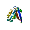
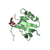
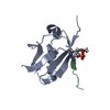
| Title | Crystal structure of K37-acetylated SUMO1 in complex with PIAS-SIM2 | |||||||||
 Components Components |
| |||||||||
 Keywords Keywords | PEPTIDE BINDING PROTEIN / SUMO1 / PIAS / SUMO INTERACTION MOTIF | |||||||||
| Function / homology |  Function and homology information Function and homology informationprotein localization to nuclear pore / SUMOylation of nuclear envelope proteins / negative regulation of transcription initiation by RNA polymerase II / SUMO is proteolytically processed / Negative regulation of activity of TFAP2 (AP-2) family transcription factors / SUMO is conjugated to E1 (UBA2:SAE1) / SUMO is transferred from E1 to E2 (UBE2I, UBC9) / negative regulation of action potential / nuclear stress granule / PML body organization ...protein localization to nuclear pore / SUMOylation of nuclear envelope proteins / negative regulation of transcription initiation by RNA polymerase II / SUMO is proteolytically processed / Negative regulation of activity of TFAP2 (AP-2) family transcription factors / SUMO is conjugated to E1 (UBA2:SAE1) / SUMO is transferred from E1 to E2 (UBE2I, UBC9) / negative regulation of action potential / nuclear stress granule / PML body organization / small protein activating enzyme binding / SUMOylation of immune response proteins / SUMOylation of SUMOylation proteins / regulation of calcium ion transmembrane transport / SUMOylation of DNA methylation proteins / Maturation of nucleoprotein / XY body / SUMOylation of RNA binding proteins / regulation of cardiac muscle cell contraction / Postmitotic nuclear pore complex (NPC) reformation / Maturation of nucleoprotein / negative regulation of protein import into nucleus / SUMOylation of ubiquitinylation proteins / ubiquitin-specific protease binding / cellular response to cadmium ion / SUMOylation of transcription factors / roof of mouth development / SUMOylation of DNA replication proteins / ubiquitin-like protein ligase binding / protein sumoylation / potassium channel regulator activity / Regulation of IFNG signaling / nuclear pore / transporter activator activity / postsynaptic cytosol / SUMOylation of DNA damage response and repair proteins / presynaptic cytosol / Transcriptional and post-translational regulation of MITF-M expression and activity / SUMOylation of transcription cofactors / SUMOylation of chromatin organization proteins / SUMOylation of intracellular receptors / regulation of protein stability / positive regulation of protein-containing complex assembly / PKR-mediated signaling / PML body / Formation of Incision Complex in GG-NER / protein tag activity / positive regulation of proteasomal ubiquitin-dependent protein catabolic process / cellular response to heat / Recruitment and ATM-mediated phosphorylation of repair and signaling proteins at DNA double strand breaks / nuclear membrane / nuclear speck / protein stabilization / nuclear body / DNA repair / negative regulation of DNA-templated transcription / ubiquitin protein ligase binding / nucleolus / glutamatergic synapse / enzyme binding / negative regulation of transcription by RNA polymerase II / RNA binding / nucleoplasm / nucleus / plasma membrane / cytosol Similarity search - Function | |||||||||
| Biological species |  Homo sapiens (human) Homo sapiens (human) | |||||||||
| Method |  X-RAY DIFFRACTION / SYNCHROTRON / MOLECULAR REPLACEMENT / Resolution: 1.549 Å X-RAY DIFFRACTION / SYNCHROTRON / MOLECULAR REPLACEMENT / Resolution: 1.549 Å | |||||||||
 Authors Authors | Lussier-Price, M. / Wahba, H.M. / Mascle, X.H. / Cappadocia, L. / Sakaguchi, K. / Omichinski, J.G. | |||||||||
| Funding support |  Canada, 2items Canada, 2items
| |||||||||
 Citation Citation | Journal: Structure / Year: 2020 Title: Characterization of a C-Terminal SUMO-Interacting Motif Present in Select PIAS-Family Proteins. Authors: Lussier-Price, M. / Mascle, X.H. / Cappadocia, L. / Kamada, R. / Sakaguchi, K. / Wahba, H.M. / Omichinski, J.G. | |||||||||
| History |
|
- Structure visualization
| Structure viewer | Molecule: MolmilJmol/JSmol |
|---|
- Downloads & links
-Download
| PDBx/mmCIF format | 6v7r.cif.gz | 120 KB | Display | PDBx/mmCIF format |
|---|---|---|---|---|
| PDB format | pdb6v7r.ent.gz | 94.4 KB | Display | PDB format |
| PDBx/mmJSON format | 6v7r.json.gz | Tree view | PDBx/mmJSON format | |
| Others |  Other downloads Other downloads |
-Validation report
| Arichive directory | https://data.pdbj.org/pub/pdb/validation_reports/v7/6v7rftp://data.pdbj.org/pub/pdb/validation_reports/v7/6v7r | HTTPS FTP |
|---|
-Related structure data
| Related structure data |  6v7pC  6v7qC  6v7sC  6uyoS C: citing same article ( S: Starting model for refinement |
|---|---|
| Similar structure data |


-Links
 PDBj
PDBj


- Assembly
| Deposited unit | 
| ||||||||
|---|---|---|---|---|---|---|---|---|---|
| 1 | 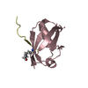
| ||||||||
| 2 | 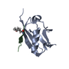
| ||||||||
| Unit cell |
|
-Components
| #1: Protein | Mass: 9567.801 Da / Num. of mol.: 2 Source method: isolated from a genetically manipulated source Source: (gene. exp.) Homo sapiens (human) / Gene: SUMO1, SMT3C, SMT3H3, UBL1, OK/SW-cl.43 / Production host:  #2: Protein/peptide | Mass: 1376.449 Da / Num. of mol.: 2 Source method: isolated from a genetically manipulated source Source: (gene. exp.) Homo sapiens (human) / Production host: #3: Water | ChemComp-HOH / |  Mass: 18.015 Da / Num. of mol.: 175 / Source method: isolated from a natural source / Formula: H2O Mass: 18.015 Da / Num. of mol.: 175 / Source method: isolated from a natural source / Formula: H2OHas ligand of interest | Y | Has protein modification | Y | |
|---|
-Experimental details
-Experiment
| Experiment | Method: X-RAY DIFFRACTION / Number of used crystals: 1 |
|---|
- Sample preparation
| Crystal | Density Matthews: 2.01 Å3/Da / Density % sol: 38.95 % |
|---|---|
| Crystal grow | Temperature: 298 K / Method: vapor diffusion, hanging drop Details: 100 mM sodium cacodylate pH 6.5, 19 to 31 % (w/v) PEG3350 and 10 mM calcium chloride. |
-Data collection
| Diffraction | Mean temperature: 100 K / Serial crystal experiment: N |
|---|---|
| Diffraction source | Source: SYNCHROTRON / Site: CHESS  / Beamline: F1 / Wavelength: 0.9775 Å / Beamline: F1 / Wavelength: 0.9775 Å |
| Detector | Type: DECTRIS PILATUS3 6M / Detector: PIXEL / Date: Mar 12, 2018 |
| Radiation | Protocol: SINGLE WAVELENGTH / Monochromatic (M) / Laue (L): M / Scattering type: x-ray |
| Radiation wavelength | Wavelength: 0.9775 Å / Relative weight: 1 |
| Reflection | Resolution: 1.549→30.263 Å / Num. obs: 24975 / % possible obs: 93.95 % / Redundancy: 5.7 % / CC1/2: 0.997 / Net I/σ(I): 12.23 |
| Reflection shell | Resolution: 1.549→1.604 Å / Num. unique obs: 1492 / CC1/2: 0.501 |
- Processing
| Software |
| |||||||||||||||||||||||||||||||||||||||||||||||||||||||||||||||||||||||||||||||||||||||||||||||||||||||||||||||||||||||||||||
|---|---|---|---|---|---|---|---|---|---|---|---|---|---|---|---|---|---|---|---|---|---|---|---|---|---|---|---|---|---|---|---|---|---|---|---|---|---|---|---|---|---|---|---|---|---|---|---|---|---|---|---|---|---|---|---|---|---|---|---|---|---|---|---|---|---|---|---|---|---|---|---|---|---|---|---|---|---|---|---|---|---|---|---|---|---|---|---|---|---|---|---|---|---|---|---|---|---|---|---|---|---|---|---|---|---|---|---|---|---|---|---|---|---|---|---|---|---|---|---|---|---|---|---|---|---|---|
| Refinement | Method to determine structure: MOLECULAR REPLACEMENT Starting model: 6UYO Resolution: 1.549→30.263 Å / SU ML: 0.22 / Cross valid method: THROUGHOUT / σ(F): 1.34 / Phase error: 25.1
| |||||||||||||||||||||||||||||||||||||||||||||||||||||||||||||||||||||||||||||||||||||||||||||||||||||||||||||||||||||||||||||
| Solvent computation | Shrinkage radii: 0.9 Å / VDW probe radii: 1.11 Å | |||||||||||||||||||||||||||||||||||||||||||||||||||||||||||||||||||||||||||||||||||||||||||||||||||||||||||||||||||||||||||||
| Displacement parameters | Biso max: 158.86 Å2 / Biso mean: 35.1703 Å2 / Biso min: 15.89 Å2 | |||||||||||||||||||||||||||||||||||||||||||||||||||||||||||||||||||||||||||||||||||||||||||||||||||||||||||||||||||||||||||||
| Refinement step | Cycle: final / Resolution: 1.549→30.263 Å
| |||||||||||||||||||||||||||||||||||||||||||||||||||||||||||||||||||||||||||||||||||||||||||||||||||||||||||||||||||||||||||||
| LS refinement shell | Refine-ID: X-RAY DIFFRACTION / Rfactor Rfree error: 0
| |||||||||||||||||||||||||||||||||||||||||||||||||||||||||||||||||||||||||||||||||||||||||||||||||||||||||||||||||||||||||||||
| Refinement TLS params. | Method: refined / Refine-ID: X-RAY DIFFRACTION
| |||||||||||||||||||||||||||||||||||||||||||||||||||||||||||||||||||||||||||||||||||||||||||||||||||||||||||||||||||||||||||||
| Refinement TLS group |
|
