 Movie
Movie Controller


+ Open data

- Basic information
| Entry | Database: PDB / ID: 6qiy | ||||||
|---|---|---|---|---|---|---|---|
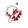
| Title | CI-2, conformation 1 | ||||||
 Components Components | Subtilisin-chymotrypsin inhibitor-2A | ||||||
 Keywords Keywords | PLANT PROTEIN / CHYMOTRYPSIN INHIBITOR 2 / protease inhibitor | ||||||
| Function / homology |  Function and homology information Function and homology information | ||||||
| Biological species |  | ||||||
| Method |  X-RAY DIFFRACTION / SYNCHROTRON / MOLECULAR REPLACEMENT / Resolution: 1.5 Å X-RAY DIFFRACTION / SYNCHROTRON / MOLECULAR REPLACEMENT / Resolution: 1.5 Å | ||||||
 Authors Authors | Romero, A. / Ruiz, F.M. | ||||||
 Citation Citation | Journal: Nat Commun / Year: 2019 Title: Engineering protein assemblies with allosteric control via monomer fold-switching. Authors: Luis A Campos / Rajendra Sharma / Sara Alvira / Federico M Ruiz / Beatriz Ibarra-Molero / Mourad Sadqi / Carlos Alfonso / Germán Rivas / Jose M Sanchez-Ruiz / Antonio Romero Garrido / José ...Authors: Luis A Campos / Rajendra Sharma / Sara Alvira / Federico M Ruiz / Beatriz Ibarra-Molero / Mourad Sadqi / Carlos Alfonso / Germán Rivas / Jose M Sanchez-Ruiz / Antonio Romero Garrido / José M Valpuesta / Victor Muñoz /    Abstract: The macromolecular machines of life use allosteric control to self-assemble, dissociate and change shape in response to signals. Despite enormous interest, the design of nanoscale allosteric ...The macromolecular machines of life use allosteric control to self-assemble, dissociate and change shape in response to signals. Despite enormous interest, the design of nanoscale allosteric assemblies has proven tremendously challenging. Here we present a proof of concept of allosteric assembly in which an engineered fold switch on the protein monomer triggers or blocks assembly. Our design is based on the hyper-stable, naturally monomeric protein CI2, a paradigm of simple two-state folding, and the toroidal arrangement with 6-fold symmetry that it only adopts in crystalline form. We engineer CI2 to enable a switch between the native and an alternate, latent fold that self-assembles onto hexagonal toroidal particles by exposing a favorable inter-monomer interface. The assembly is controlled on demand via the competing effects of temperature and a designed short peptide. These findings unveil a remarkable potential for structural metamorphosis in proteins and demonstrate key principles for engineering protein-based nanomachinery. | ||||||
| History |
|
- Structure visualization
| Structure viewer | Molecule: MolmilJmol/JSmol |
|---|
- Downloads & links
-Download
| PDBx/mmCIF format | 6qiy.cif.gz | 41.5 KB | Display | PDBx/mmCIF format |
|---|---|---|---|---|
| PDB format | pdb6qiy.ent.gz | 28.2 KB | Display | PDB format |
| PDBx/mmJSON format | 6qiy.json.gz | Tree view | PDBx/mmJSON format | |
| Others |  Other downloads Other downloads |
-Validation report
| Arichive directory | https://data.pdbj.org/pub/pdb/validation_reports/qi/6qiyftp://data.pdbj.org/pub/pdb/validation_reports/qi/6qiy | HTTPS FTP |
|---|
-Related structure data
| Related structure data | 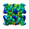 4568C 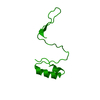 6qizC  2ci2S S: Starting model for refinement C: citing same article ( |
|---|---|
| Similar structure data |


-Links
 PDBj
PDBj- Assembly
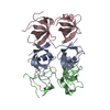
| Deposited unit | 
| ||||||||
|---|---|---|---|---|---|---|---|---|---|
| 1 | x 12
| ||||||||
| Unit cell |
| ||||||||
| Components on special symmetry positions |
|
-Components
| #1: Protein | Mass: 7411.657 Da / Num. of mol.: 1 / Fragment: UNP RESIDUES 20-84 Source method: isolated from a genetically manipulated source Source: (gene. exp.)  |
|---|---|
| #2: Water | ChemComp-HOH /  Mass: 18.015 Da / Num. of mol.: 65 / Source method: isolated from a natural source / Formula: H2O Mass: 18.015 Da / Num. of mol.: 65 / Source method: isolated from a natural source / Formula: H2O |
-Experimental details
-Experiment
| Experiment | Method: X-RAY DIFFRACTION / Number of used crystals: 1 |
|---|
- Sample preparation
| Crystal | Density Matthews: 2.45 Å3/Da / Density % sol: 49.76 % |
|---|---|
| Crystal grow | Temperature: 293 K / Method: vapor diffusion, sitting drop / pH: 8.5 Details: 1.4 M AMMONIUM SULPHATE AND 100 MM TRIS-HCL PH 8.5, VAPOR DIFFUSION, SITTING DROP, TEMPERATURE 293K PH range: 8.5 |
-Data collection
| Diffraction | Mean temperature: 100 K / Serial crystal experiment: N |
|---|---|
| Diffraction source | Source: SYNCHROTRON / Site: ALBA / Beamline: XALOC / Wavelength: 1 Å |
| Detector | Type: DECTRIS PILATUS3 S 6M / Detector: PIXEL / Date: Mar 24, 2015 |
| Radiation | Protocol: SINGLE WAVELENGTH / Monochromatic (M) / Laue (L): M / Scattering type: x-ray |
| Radiation wavelength | Wavelength: 1 Å / Relative weight: 1 |
| Reflection | Resolution: 1.5→38.55 Å / Num. obs: 11793 / % possible obs: 100 % / Redundancy: 18.1 % / Biso Wilson estimate: 22.61 Å2 / Rmerge(I) obs: 0.059 / Rsym value: 0.02 / Net I/σ(I): 23 |
| Reflection shell | Resolution: 1.5→1.55 Å / Mean I/σ(I) obs: 1.8 / % possible all: 100 |
- Processing
| Software |
| ||||||||||||||||||||||||||||||||||||||||||||||||||||||||||||||||||||||||||||||||||||||||||||||||||||
|---|---|---|---|---|---|---|---|---|---|---|---|---|---|---|---|---|---|---|---|---|---|---|---|---|---|---|---|---|---|---|---|---|---|---|---|---|---|---|---|---|---|---|---|---|---|---|---|---|---|---|---|---|---|---|---|---|---|---|---|---|---|---|---|---|---|---|---|---|---|---|---|---|---|---|---|---|---|---|---|---|---|---|---|---|---|---|---|---|---|---|---|---|---|---|---|---|---|---|---|---|---|
| Refinement | Method to determine structure: MOLECULAR REPLACEMENT Starting model: 2CI2 Resolution: 1.5→38.55 Å / SU ML: 0.17 / Cross valid method: FREE R-VALUE / σ(F): 1.33 / Phase error: 37.6
| ||||||||||||||||||||||||||||||||||||||||||||||||||||||||||||||||||||||||||||||||||||||||||||||||||||
| Solvent computation | Shrinkage radii: 0.9 Å / VDW probe radii: 1.11 Å | ||||||||||||||||||||||||||||||||||||||||||||||||||||||||||||||||||||||||||||||||||||||||||||||||||||
| Refinement step | Cycle: LAST / Resolution: 1.5→38.55 Å
| ||||||||||||||||||||||||||||||||||||||||||||||||||||||||||||||||||||||||||||||||||||||||||||||||||||
| Refine LS restraints |
| ||||||||||||||||||||||||||||||||||||||||||||||||||||||||||||||||||||||||||||||||||||||||||||||||||||
| LS refinement shell |
| ||||||||||||||||||||||||||||||||||||||||||||||||||||||||||||||||||||||||||||||||||||||||||||||||||||
| Refinement TLS params. | Method: refined / Refine-ID: X-RAY DIFFRACTION
| ||||||||||||||||||||||||||||||||||||||||||||||||||||||||||||||||||||||||||||||||||||||||||||||||||||
| Refinement TLS group |
|
