[English] 日本語
Yorodumi
- PDB-2wfw: Structure and activity of the N-terminal substrate recognition do... -

+
Open data


ID or keywords:

Loading...

-
Basic information

Entry
Database: PDB / ID: 2wfw
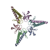
Title
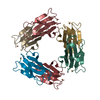
Structure and activity of the N-terminal substrate recognition domains in proteasomal ATPases - The Arc domain structure
ComponentsARC
KeywordsATP-BINDING PROTEIN / PROTEASOMAL ATPASES / PAN / ARC / AAA / ATP-BINDING / NUCLEOTIDE-BINDING
Function / homology
Function and homology information


proteasome-activating nucleotidase complex / CTPase activity / proteasome complex / proteasomal protein catabolic process / modification-dependent protein catabolic process / ATP hydrolysis activity / ATP binding
Similarity search - Function
Proteasome ATPase / Proteasomal ATPase, N-terminal OB domain / Proteasomal ATPase OB N-terminal domain / Proteasomal ATPase OB C-terminal domain / Proteasomal ATPase OB C-terminal domain / ATPase, AAA-type, conserved site / AAA-protein family signature. / Nucleic acid-binding proteins / ATPase family associated with various cellular activities (AAA) / ATPase, AAA-type, core ...Proteasome ATPase / Proteasomal ATPase, N-terminal OB domain / Proteasomal ATPase OB N-terminal domain / Proteasomal ATPase OB C-terminal domain / Proteasomal ATPase OB C-terminal domain / ATPase, AAA-type, conserved site / AAA-protein family signature. / Nucleic acid-binding proteins / ATPase family associated with various cellular activities (AAA) / ATPase, AAA-type, core / OB fold (Dihydrolipoamide Acetyltransferase, E2P) / ATPases associated with a variety of cellular activities / AAA+ ATPase domain / Beta Barrel / P-loop containing nucleoside triphosphate hydrolase / Mainly Beta
Similarity search - Domain/homology
Proteasome-associated ATPase
Similarity search - Component
Biological speciesRHODOCOCCUS ERYTHROPOLIS (bacteria)
MethodX-RAY DIFFRACTION / SYNCHROTRON / MIR / Resolution: 1.6 Å
AuthorsDjuranovic, S. / Hartmann, M.D. / Habeck, M. / Ursinus, A. / Zwickl, P. / Martin, J. / Lupas, A.N. / Zeth, K.
CitationJournal: Mol.Cell / Year: 2009
Title: Structure and Activity of the N-Terminal Substrate Recognition Domains in Proteasomal Atpases.
Authors: Djuranovic, S. / Hartmann, M.D. / Habeck, M. / Ursinus, A. / Zwickl, P. / Martin, J. / Lupas, A.N. / Zeth, K.
History
DepositionApr 15, 2009Deposition site: PDBE / Processing site: PDBE
Revision 1.0May 12, 2009Provider: repository / Type: Initial release
Revision 1.1Jul 13, 2011Group: Advisory / Version format compliance
Revision 1.2May 8, 2024Group: Data collection / Database references ...Data collection / Database references / Other / Refinement description
Category: chem_comp_atom / chem_comp_bond ...chem_comp_atom / chem_comp_bond / database_2 / pdbx_database_status / struct_ncs_dom_lim
Item: _database_2.pdbx_DOI / _database_2.pdbx_database_accession ..._database_2.pdbx_DOI / _database_2.pdbx_database_accession / _pdbx_database_status.status_code_sf / _struct_ncs_dom_lim.beg_auth_comp_id / _struct_ncs_dom_lim.beg_label_asym_id / _struct_ncs_dom_lim.beg_label_comp_id / _struct_ncs_dom_lim.beg_label_seq_id / _struct_ncs_dom_lim.end_auth_comp_id / _struct_ncs_dom_lim.end_label_asym_id / _struct_ncs_dom_lim.end_label_comp_id / _struct_ncs_dom_lim.end_label_seq_id
Remark 700 SHEET DETERMINATION METHOD: DSSP THE SHEETS PRESENTED AS "AB" IN EACH CHAIN ON SHEET RECORDS BELOW ... SHEET DETERMINATION METHOD: DSSP THE SHEETS PRESENTED AS "AB" IN EACH CHAIN ON SHEET RECORDS BELOW IS ACTUALLY AN 15-STRANDED BARREL THIS IS REPRESENTED BY A 16-STRANDED SHEET IN WHICH THE FIRST AND LAST STRANDS ARE IDENTICAL.

-
Structure visualization

Structure viewerMolecule:
MolmilJmol/JSmol

Downloads & links

-
Assembly

Deposited unit
A: ARC
B: ARC
C: ARC


Theoretical massNumber of molelcules
Total (without water)49,8233
Polymers49,8233
Non-polymers00
Water8,305461
1
A: ARC
B: ARC
C: ARC

A: ARC
B: ARC
C: ARC


Theoretical massNumber of molelcules
Total (without water)99,6466
Polymers99,6466
Non-polymers00
Water1086
TypeNameSymmetry operationNumber
identity operation1_555x,y,z1
crystal symmetry operation5_555x-y,-y,-z+1/31
Buried area10210 Å2
ΔGint-28.18 kcal/mol
Surface area34740 Å2
MethodPISA
Unit cell
Length a, b, c (Å)100.330, 100.330, 88.000
Angle α, β, γ (deg.)90.00, 90.00, 120.00
Int Tables number154
Space group name H-MP3221
Noncrystallographic symmetry (NCS)NCS domain:
IDEns-IDDetails
11A
21B
31C

NCS domain segments:

Component-ID: 1 / Ens-ID: 1 / Beg auth comp-ID: SER / Beg label comp-ID: SER / End auth comp-ID: GLY / End label comp-ID: GLY / Refine code: 6 / Auth seq-ID: 81 - 160 / Label seq-ID: 8 - 87

Dom-IDAuth asym-IDLabel asym-ID
1AA
2BB
3CC

-
Components

#1: Protein ARC


Mass: 16607.674 Da / Num. of mol.: 3 / Fragment: RESIDUES 73-225
Source method: isolated from a genetically manipulated source
Source: (gene. exp.) RHODOCOCCUS ERYTHROPOLIS (bacteria) / Plasmid: PET30B (NOVAGEN) / Production host: ESCHERICHIA COLI (E. coli) / Strain (production host): BL21(DE3) / References: UniProt: O50202
#2: Water ChemComp-HOH / water


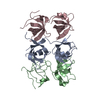
Mass: 18.015 Da / Num. of mol.: 461 / Source method: isolated from a natural source / Formula: H2O

-
Experimental details

-
Experiment

ExperimentMethod: X-RAY DIFFRACTION / Number of used crystals: 1

-
Sample preparation

CrystalDensity Matthews: 2.7 Å3/Da / Density % sol: 55 % / Description: NONE
Crystal growpH: 6.5 / Details: 0.05 MM CACL2, 0.1 M BIS-TRIS PH 6.5, 30% PEG550

-
Data collection

DiffractionMean temperature: 100 K
Diffraction sourceSource: SYNCHROTRON / Site: SLS / Beamline: X06SA / Wavelength: 1
DetectorType: MARRESEARCH / Detector: CCD
RadiationProtocol: SINGLE WAVELENGTH / Monochromatic (M) / Laue (L): M / Scattering type: x-ray
Radiation wavelengthWavelength: 1 Å / Relative weight: 1
ReflectionResolution: 1.6→30 Å / Num. obs: 64443 / % possible obs: 95.2 % / Observed criterion σ(I): 3 / Redundancy: 7 % / Biso Wilson estimate: 28 Å2 / Rmerge(I) obs: 0.07 / Net I/σ(I): 14.7
Reflection shellResolution: 1.6→1.7 Å / Redundancy: 6.7 % / Rmerge(I) obs: 0.53 / Mean I/σ(I) obs: 3.2 / % possible all: 78.6

-
Processing

Software
NameVersionClassification
REFMAC5.2.0019refinement
XDSdata reduction
XDSdata scaling
SHARPphasing
RefinementMethod to determine structure: MIR
Starting model: NONE

Resolution: 1.6→20 Å / Cor.coef. Fo:Fc: 0.944 / Cor.coef. Fo:Fc free: 0.922 / SU B: 2.843 / SU ML: 0.053 / TLS residual ADP flag: LIKELY RESIDUAL / Cross valid method: THROUGHOUT / ESU R: 0.089 / ESU R Free: 0.093 / Stereochemistry target values: MAXIMUM LIKELIHOOD / Details: HYDROGENS HAVE BEEN ADDED IN THE RIDING POSITIONS.
RfactorNum. reflection% reflectionSelection details
Rfree0.24641 3222 5 %RANDOM
Rwork0.20825 ---
obs0.21011 61206 100 %-
Solvent computationIon probe radii: 0.8 Å / Shrinkage radii: 0.8 Å / VDW probe radii: 1.4 Å / Solvent model: MASK
Displacement parametersBiso mean: 26.377 Å2
Baniso -1Baniso -2Baniso -3
1-0.18 Å20.09 Å20 Å2
2--0.18 Å20 Å2
3----0.28 Å2
Refinement stepCycle: LAST / Resolution: 1.6→20 Å
ProteinNucleic acidLigandSolventTotal
Num. atoms2807 0 0 461 3268
Refine LS restraints
Refine-IDTypeDev idealDev ideal targetNumber
X-RAY DIFFRACTIONr_bond_refined_d0.0140.0222881
X-RAY DIFFRACTIONr_bond_other_d
X-RAY DIFFRACTIONr_angle_refined_deg1.6341.983926
X-RAY DIFFRACTIONr_angle_other_deg
X-RAY DIFFRACTIONr_dihedral_angle_1_deg7.415381
X-RAY DIFFRACTIONr_dihedral_angle_2_deg26.26723.898118
X-RAY DIFFRACTIONr_dihedral_angle_3_deg13.2615477
X-RAY DIFFRACTIONr_dihedral_angle_4_deg15.3611523
X-RAY DIFFRACTIONr_chiral_restr0.1370.2481
X-RAY DIFFRACTIONr_gen_planes_refined0.0060.022129
X-RAY DIFFRACTIONr_gen_planes_other
X-RAY DIFFRACTIONr_nbd_refined0.2360.21361
X-RAY DIFFRACTIONr_nbd_other
X-RAY DIFFRACTIONr_nbtor_refined0.3050.22000
X-RAY DIFFRACTIONr_nbtor_other
X-RAY DIFFRACTIONr_xyhbond_nbd_refined0.1460.2329
X-RAY DIFFRACTIONr_xyhbond_nbd_other
X-RAY DIFFRACTIONr_metal_ion_refined
X-RAY DIFFRACTIONr_metal_ion_other
X-RAY DIFFRACTIONr_symmetry_vdw_refined0.2540.283
X-RAY DIFFRACTIONr_symmetry_vdw_other
X-RAY DIFFRACTIONr_symmetry_hbond_refined0.1550.230
X-RAY DIFFRACTIONr_symmetry_hbond_other
X-RAY DIFFRACTIONr_symmetry_metal_ion_refined
X-RAY DIFFRACTIONr_symmetry_metal_ion_other
X-RAY DIFFRACTIONr_mcbond_it1.0731.51915
X-RAY DIFFRACTIONr_mcbond_other
X-RAY DIFFRACTIONr_mcangle_it1.71522998
X-RAY DIFFRACTIONr_mcangle_other
X-RAY DIFFRACTIONr_scbond_it2.44231075
X-RAY DIFFRACTIONr_scbond_other
X-RAY DIFFRACTIONr_scangle_it3.7464.5921
X-RAY DIFFRACTIONr_scangle_other
X-RAY DIFFRACTIONr_long_range_B_refined
X-RAY DIFFRACTIONr_long_range_B_other
X-RAY DIFFRACTIONr_rigid_bond_restr
X-RAY DIFFRACTIONr_sphericity_free
X-RAY DIFFRACTIONr_sphericity_bonded
Refine LS restraints NCS

Ens-ID: 1 / Number: 585 / Refine-ID: X-RAY DIFFRACTION

Dom-IDAuth asym-IDTypeRms dev position (Å)Weight position
1Aloose positional0.45
2Bloose positional0.365
3Cloose positional0.545
1Aloose thermal1.5310
2Bloose thermal1.3110
3Cloose thermal1.2810
LS refinement shellResolution: 1.6→1.642 Å / Total num. of bins used: 20
RfactorNum. reflection% reflection
Rfree0.299 132 -
Rwork0.287 2491 -
obs--100 %
Refinement TLS params.

Method: refined / Refine-ID: X-RAY DIFFRACTION

IDL112)L122)L132)L222)L232)L332)S11 (Å °)S12 (Å °)S13 (Å °)S21 (Å °)S22 (Å °)S23 (Å °)S31 (Å °)S32 (Å °)S33 (Å °)T112)T122)T132)T222)T232)T332)Origin x (Å)Origin y (Å)Origin z (Å)
11.5791-0.1116-0.2661.5829-0.39811.3246-0.02040.00070.00290.0590.06620.0533-0.11440.0037-0.04590.0079-0.00230.018-0.02740.0176-0.071852.77522.63116.578
22.9773-0.573-0.41662.7898-2.14552.2939-0.1883-0.0728-0.13410.3179-0.0681-0.1207-0.15210.23390.2564-0.0014-0.0711-0.08090.02670.0249-0.074272.49319.31620.667
31.35270.1609-0.81922.64190.8662.9217-0.0448-0.1588-0.16110.1892-0.0719-0.29430.01730.33050.1166-0.0683-0.055-0.0444-0.00420.0489-0.041874.76622.03711.502
44.1721.16851.02383.1407-0.63234.19550.0457-0.0687-0.09330.3923-0.225-0.3097-0.30090.44510.17930.0187-0.0964-0.07760.04840.0633-0.039774.20920.43220.548
51.19680.17310.51761.8866-0.08081.1171-0.01520.02660.0152-0.19660.04050.09070.0702-0.0365-0.02530.0147-0.0497-0.0056-0.01960.006-0.071755.85913.393-3.458
63.9321-0.2767-0.15247.43651.40642.00820.08510.02030.0974-0.44310.117-0.7608-0.04030.2568-0.2022-0.0705-0.02670.0985-0.0014-0.0018-0.015575.258.083-5.69
73.38230.60480.14085.49661.21732.17880.0278-0.10580.0369-0.13750.0152-0.1784-0.04960.0733-0.043-0.0516-0.04310.04360.00240.0019-0.034871.37113.253-0.578
81.7259-0.8041-0.3710.8832-0.36550.6495-0.0216-0.0885-0.04260.20980.0467-0.0137-0.12990.0316-0.02510.07450.0452-0.007-0.0153-0.0022-0.077656.468.8535.735
93.42840.9258-2.26322.1913-3.04736.6671-0.0188-0.01420.10730.2833-0.2724-0.4274-0.64670.57760.2912-0.0035-0.0821-0.10230.02320.0444-0.048275.1213.20531.297
104.5939-0.83530.72753.7645-1.51523.48160.07310.0778-0.01060.3058-0.1832-0.2325-0.23320.18010.1102-0.0038-0.0063-0.0678-0.00520.0319-0.05771.4646.32833.204
Refinement TLS group
IDRefine-IDRefine TLS-IDAuth asym-IDAuth seq-ID
1X-RAY DIFFRACTION1A80 - 135
2X-RAY DIFFRACTION2A136 - 147
3X-RAY DIFFRACTION3A148 - 176
4X-RAY DIFFRACTION4A200 - 220
5X-RAY DIFFRACTION5B80 - 146
6X-RAY DIFFRACTION6B147 - 174
7X-RAY DIFFRACTION7B200 - 214
8X-RAY DIFFRACTION8C80 - 146
9X-RAY DIFFRACTION9C147 - 175
10X-RAY DIFFRACTION10C200 - 214

+
About Yorodumi

-
News

-
Feb 9, 2022. New format data for meta-information of EMDB entries

New format data for meta-information of EMDB entries

  • Version 3 of the EMDB header file is now the official format.
  • The previous official version 1.9 will be removed from the archive.

Related info.:EMDB header

External links:wwPDB to switch to version 3 of the EMDB data model

-
Aug 12, 2020. Covid-19 info

Covid-19 info

URL: https://pdbjlvh1.pdbj.org/emnavi/covid19.php

New page: Covid-19 featured information page in EM Navigator.

Related info.:Covid-19 info / Mar 5, 2020. Novel coronavirus structure data

+
Mar 5, 2020. Novel coronavirus structure data

Novel coronavirus structure data

Related info.:Yorodumi Speices / Aug 12, 2020. Covid-19 info

External links:COVID-19 featured content - PDBj / Molecule of the Month (242):Coronavirus Proteases

+
Jan 31, 2019. EMDB accession codes are about to change! (news from PDBe EMDB page)

EMDB accession codes are about to change! (news from PDBe EMDB page)

  • The allocation of 4 digits for EMDB accession codes will soon come to an end. Whilst these codes will remain in use, new EMDB accession codes will include an additional digit and will expand incrementally as the available range of codes is exhausted. The current 4-digit format prefixed with “EMD-” (i.e. EMD-XXXX) will advance to a 5-digit format (i.e. EMD-XXXXX), and so on. It is currently estimated that the 4-digit codes will be depleted around Spring 2019, at which point the 5-digit format will come into force.
  • The EM Navigator/Yorodumi systems omit the EMD- prefix.

Related info.:Q: What is EMD? / ID/Accession-code notation in Yorodumi/EM Navigator

External links:EMDB Accession Codes are Changing Soon! / Contact to PDBj

+
Jul 12, 2017. Major update of PDB

Major update of PDB

  • wwPDB released updated PDB data conforming to the new PDBx/mmCIF dictionary.
  • This is a major update changing the version number from 4 to 5, and with Remediation, in which all the entries are updated.
  • In this update, many items about electron microscopy experimental information are reorganized (e.g. em_software).
  • Now, EM Navigator and Yorodumi are based on the updated data.

External links:wwPDB Remediation / Enriched Model Files Conforming to OneDep Data Standards Now Available in the PDB FTP Archive

-
Yorodumi

Thousand views of thousand structures

  • Yorodumi is a browser for structure data from EMDB, PDB, SASBDB, etc.
  • This page is also the successor to EM Navigator detail page, and also detail information page/front-end page for Omokage search.
  • The word "yorodu" (or yorozu) is an old Japanese word meaning "ten thousand". "mi" (miru) is to see.

Related info.:EMDB / PDB / SASBDB / Comparison of 3 databanks / Yorodumi Search / Aug 31, 2016. New EM Navigator & Yorodumi / Yorodumi Papers / Jmol/JSmol / Function and homology information / Changes in new EM Navigator and Yorodumi

Read more