[English] 日本語
Yorodumi
- PDB-3fp9: Crystal structure of Intern Domain of proteasome-associated ATPas... -

+
Open data


ID or keywords:

Loading...

-
Basic information

Entry
Database: PDB / ID: 3fp9
TitleCrystal structure of Intern Domain of proteasome-associated ATPase, Mycobacterium tuberculosis
ComponentsProteasome-associated ATPase
KeywordsHYDROLASE / five strand beta barrel / hexamer / ATP-binding / Nucleotide-binding / AAA ATPase / Proteasomal ATPase
Function / homology
Function and homology information


ubiquitin-like protein reader activity / symbiont defense to host-produced reactive oxygen species / proteasome-activating nucleotidase complex / symbiont-mediated perturbation of host defenses / response to nitrosative stress / cell wall / ATP-dependent peptidase activity / proteasomal ubiquitin-independent protein catabolic process / protein unfolding / cellular response to nitric oxide ...ubiquitin-like protein reader activity / symbiont defense to host-produced reactive oxygen species / proteasome-activating nucleotidase complex / symbiont-mediated perturbation of host defenses / response to nitrosative stress / cell wall / ATP-dependent peptidase activity / proteasomal ubiquitin-independent protein catabolic process / protein unfolding / cellular response to nitric oxide / peptidoglycan-based cell wall / proteasomal protein catabolic process / modification-dependent protein catabolic process / ATP hydrolysis activity / ATP binding / identical protein binding / plasma membrane
Similarity search - Function
Proteasome ATPase / Proteasomal ATPase, N-terminal OB domain / Proteasomal ATPase OB N-terminal domain / Proteasomal ATPase OB C-terminal domain / Proteasomal ATPase OB C-terminal domain / ATPase, AAA-type, conserved site / AAA-protein family signature. / Nucleic acid-binding proteins / ATPase family associated with various cellular activities (AAA) / ATPase, AAA-type, core ...Proteasome ATPase / Proteasomal ATPase, N-terminal OB domain / Proteasomal ATPase OB N-terminal domain / Proteasomal ATPase OB C-terminal domain / Proteasomal ATPase OB C-terminal domain / ATPase, AAA-type, conserved site / AAA-protein family signature. / Nucleic acid-binding proteins / ATPase family associated with various cellular activities (AAA) / ATPase, AAA-type, core / OB fold (Dihydrolipoamide Acetyltransferase, E2P) / ATPases associated with a variety of cellular activities / AAA+ ATPase domain / Nucleic acid-binding, OB-fold / Beta Barrel / P-loop containing nucleoside triphosphate hydrolase / Mainly Beta
Similarity search - Domain/homology
Proteasome-associated ATPase / Proteasome-associated ATPase
Similarity search - Component
Biological speciesMycobacterium tuberculosis (bacteria)
MethodX-RAY DIFFRACTION / SYNCHROTRON / SIR/AS, molecular replacement / Resolution: 2 Å
AuthorsLi, H. / Wang, T.
CitationJournal: Structure / Year: 2009
Title: Structural Insights on the Mycobacterium tuberculosis Proteasomal ATPase Mpa.
Authors: Wang, T. / Li, H. / Lin, G. / Tang, C. / Li, D. / Nathan, C. / Darwin, K.H. / Li, H.
History
DepositionJan 4, 2009Deposition site: RCSB / Processing site: RCSB
Revision 1.0Oct 27, 2009Provider: repository / Type: Initial release
Revision 1.1Jul 13, 2011Group: Version format compliance
Revision 1.2Feb 21, 2024Group: Data collection / Database references
Category: chem_comp_atom / chem_comp_bond ...chem_comp_atom / chem_comp_bond / database_2 / struct_ref_seq_dif
Item: _database_2.pdbx_DOI / _database_2.pdbx_database_accession / _struct_ref_seq_dif.details

-
Structure visualization

Structure viewerMolecule:
MolmilJmol/JSmol

Downloads & links

-
Assembly

Deposited unit
A: Proteasome-associated ATPase
B: Proteasome-associated ATPase
C: Proteasome-associated ATPase
D: Proteasome-associated ATPase
E: Proteasome-associated ATPase
F: Proteasome-associated ATPase
G: Proteasome-associated ATPase
H: Proteasome-associated ATPase
I: Proteasome-associated ATPase
J: Proteasome-associated ATPase
K: Proteasome-associated ATPase
L: Proteasome-associated ATPase


Theoretical massNumber of molelcules
Total (without water)200,13412
Polymers200,13412
Non-polymers00
Water15,601866
1


  • Idetical with deposited unit
  • defined by software
TypeNameSymmetry operationNumber
identity operation1_555x,y,z1
Buried area28260 Å2
ΔGint-113 kcal/mol
Surface area79460 Å2
MethodPISA
2
A: Proteasome-associated ATPase
B: Proteasome-associated ATPase
C: Proteasome-associated ATPase
D: Proteasome-associated ATPase
E: Proteasome-associated ATPase
F: Proteasome-associated ATPase


Theoretical massNumber of molelcules
Total (without water)100,0676
Polymers100,0676
Non-polymers00
Water1086
TypeNameSymmetry operationNumber
identity operation1_555x,y,z1
Buried area12940 Å2
ΔGint-39 kcal/mol
Surface area41100 Å2
MethodPISA
3
G: Proteasome-associated ATPase
H: Proteasome-associated ATPase
I: Proteasome-associated ATPase
J: Proteasome-associated ATPase
K: Proteasome-associated ATPase
L: Proteasome-associated ATPase


Theoretical massNumber of molelcules
Total (without water)100,0676
Polymers100,0676
Non-polymers00
Water1086
TypeNameSymmetry operationNumber
identity operation1_555x,y,z1
Buried area12840 Å2
ΔGint-38 kcal/mol
Surface area40840 Å2
MethodPISA
Unit cell
Length a, b, c (Å)74.860, 74.994, 200.846
Angle α, β, γ (deg.)90.00, 90.32, 90.00
Int Tables number4
Space group name H-MP1211
Details
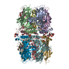
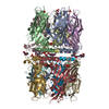
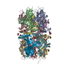
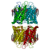
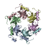
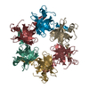
the biological assembly is a hexamer, which is composed of six monomer of Inter-Domain and contained in the asymmetric unit, two biological hexamer form crystal packing.

-
Components

#1: Protein
Proteasome-associated ATPase


Mass: 16677.811 Da / Num. of mol.: 12 / Fragment: Intern domain
Source method: isolated from a genetically manipulated source
Source: (gene. exp.) Mycobacterium tuberculosis (bacteria) / Production host: Escherichia coli (E. coli) / References: UniProt: P63345, UniProt: P9WQN5*PLUS
#2: Water ChemComp-HOH / water / Water


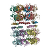
Mass: 18.015 Da / Num. of mol.: 866 / Source method: isolated from a natural source / Formula: H2O

-
Experimental details

-
Experiment

ExperimentMethod: X-RAY DIFFRACTION / Number of used crystals: 3

-
Sample preparation

CrystalDensity Matthews: 2.87 Å3/Da / Density % sol: 57.09 %
Crystal growTemperature: 294 K / Method: vapor diffusion, hanging drop / pH: 6
Details: 20% PEG2000, 50 mM MgCl2, 50 mM NaCl, 0.1M MES, C-HEGA, pH 6.0, VAPOR DIFFUSION, HANGING DROP, temperature 294K

-
Data collection

Diffraction
IDMean temperature (K)Crystal-ID
11001
21001
31001
41001
Diffraction source
SourceSiteBeamlineIDWavelength (Å)
SYNCHROTRONNSLS X2511
SYNCHROTRONNSLS X29A21
SYNCHROTRONNSLS X2531
SYNCHROTRONNSLS X29A41.0066
Detector
TypeIDDetectorDate
ADSC QUANTUM 3151CCDNov 24, 2008
ADSC QUANTUM 3152CCDOct 27, 2008
ADSC QUANTUM 3153CCDSep 26, 2008
ADSC QUANTUM 3154CCDAug 19, 2008
Radiation
IDMonochromatorProtocolMonochromatic (M) / Laue (L)Scattering typeWavelength-ID
1Si-111SINGLE WAVELENGTHMx-ray1
2Si-111SINGLE WAVELENGTHx-ray1
3Si-111SINGLE WAVELENGTHx-ray1
4Si-111SINGLE WAVELENGTHx-ray2
Radiation wavelength
IDWavelength (Å)Relative weight
111
21.00661
ReflectionResolution: 2→20 Å / Num. obs: 147655 / % possible obs: 98 % / Observed criterion σ(F): 2 / Observed criterion σ(I): 1 / Redundancy: 7 % / Rmerge(I) obs: 0.19
Reflection shellResolution: 2→2.07 Å / Rmerge(I) obs: 0.5 / Mean I/σ(I) obs: 2.5 / % possible all: 89

-
Processing

Software
NameVersionClassification
CBASSdata collection
PHENIXmodel building
SHELXmodel building
SOLVEphasing
PHASERphasing
REFMAC5.2.0019refinement
HKL-2000data reduction
HKL-2000data scaling
PHENIXphasing
SHELXphasing
RefinementMethod to determine structure: SIR/AS, molecular replacement
Resolution: 2→20 Å / Cor.coef. Fo:Fc: 0.944 / Cor.coef. Fo:Fc free: 0.909 / SU B: 4.095 / SU ML: 0.117 / Cross valid method: THROUGHOUT / ESU R: 0.175 / ESU R Free: 0.171 / Stereochemistry target values: MAXIMUM LIKELIHOOD / Details: HYDROGENS HAVE BEEN ADDED IN THE RIDING POSITIONS
RfactorNum. reflection% reflectionSelection details
Rfree0.26632 7416 5 %RANDOM
Rwork0.21286 ---
obs0.21556 140155 98.13 %-
Solvent computationIon probe radii: 0.8 Å / Shrinkage radii: 0.8 Å / VDW probe radii: 1.2 Å / Solvent model: MASK
Displacement parametersBiso mean: 33.471 Å2
Baniso -1Baniso -2Baniso -3
1-0.86 Å20 Å2-0.19 Å2
2--0.92 Å20 Å2
3----1.78 Å2
Refinement stepCycle: LAST / Resolution: 2→20 Å
ProteinNucleic acidLigandSolventTotal
Num. atoms12869 0 0 866 13735
Refine LS restraints
Refine-IDTypeDev idealDev ideal targetNumber
X-RAY DIFFRACTIONr_bond_refined_d0.0180.02213048
X-RAY DIFFRACTIONr_bond_other_d
X-RAY DIFFRACTIONr_angle_refined_deg1.6821.98817712
X-RAY DIFFRACTIONr_angle_other_deg
X-RAY DIFFRACTIONr_dihedral_angle_1_deg6.68251661
X-RAY DIFFRACTIONr_dihedral_angle_2_deg28.40224.011571
X-RAY DIFFRACTIONr_dihedral_angle_3_deg14.753152226
X-RAY DIFFRACTIONr_dihedral_angle_4_deg15.1715117
X-RAY DIFFRACTIONr_chiral_restr0.1270.22136
X-RAY DIFFRACTIONr_gen_planes_refined0.0070.029669
X-RAY DIFFRACTIONr_gen_planes_other
X-RAY DIFFRACTIONr_nbd_refined0.2170.35380
X-RAY DIFFRACTIONr_nbd_other
X-RAY DIFFRACTIONr_nbtor_refined0.3120.58889
X-RAY DIFFRACTIONr_nbtor_other
X-RAY DIFFRACTIONr_xyhbond_nbd_refined0.1880.51657
X-RAY DIFFRACTIONr_xyhbond_nbd_other
X-RAY DIFFRACTIONr_metal_ion_refined
X-RAY DIFFRACTIONr_metal_ion_other
X-RAY DIFFRACTIONr_symmetry_vdw_refined0.3120.374
X-RAY DIFFRACTIONr_symmetry_vdw_other
X-RAY DIFFRACTIONr_symmetry_hbond_refined0.240.519
X-RAY DIFFRACTIONr_symmetry_hbond_other
X-RAY DIFFRACTIONr_symmetry_metal_ion_refined
X-RAY DIFFRACTIONr_symmetry_metal_ion_other
X-RAY DIFFRACTIONr_mcbond_it1.91228573
X-RAY DIFFRACTIONr_mcbond_other
X-RAY DIFFRACTIONr_mcangle_it2.962313460
X-RAY DIFFRACTIONr_scbond_it2.02724922
X-RAY DIFFRACTIONr_scangle_it3.09234252
X-RAY DIFFRACTIONr_rigid_bond_restr
X-RAY DIFFRACTIONr_sphericity_free
X-RAY DIFFRACTIONr_sphericity_bonded
LS refinement shellResolution: 2→2.051 Å / Total num. of bins used: 20
RfactorNum. reflection% reflection
Rfree0.375 471 -
Rwork0.258 8814 -
obs--85.06 %

+
About Yorodumi

-
News

-
Feb 9, 2022. New format data for meta-information of EMDB entries

New format data for meta-information of EMDB entries

  • Version 3 of the EMDB header file is now the official format.
  • The previous official version 1.9 will be removed from the archive.

Related info.:EMDB header

External links:wwPDB to switch to version 3 of the EMDB data model

-
Aug 12, 2020. Covid-19 info

Covid-19 info

URL: https://pdbj.org/emnavi/covid19.php

New page: Covid-19 featured information page in EM Navigator.

Related info.:Covid-19 info / Mar 5, 2020. Novel coronavirus structure data

+
Mar 5, 2020. Novel coronavirus structure data

Novel coronavirus structure data

Related info.:Yorodumi Speices / Aug 12, 2020. Covid-19 info

External links:COVID-19 featured content - PDBj / Molecule of the Month (242):Coronavirus Proteases

+
Jan 31, 2019. EMDB accession codes are about to change! (news from PDBe EMDB page)

EMDB accession codes are about to change! (news from PDBe EMDB page)

  • The allocation of 4 digits for EMDB accession codes will soon come to an end. Whilst these codes will remain in use, new EMDB accession codes will include an additional digit and will expand incrementally as the available range of codes is exhausted. The current 4-digit format prefixed with “EMD-” (i.e. EMD-XXXX) will advance to a 5-digit format (i.e. EMD-XXXXX), and so on. It is currently estimated that the 4-digit codes will be depleted around Spring 2019, at which point the 5-digit format will come into force.
  • The EM Navigator/Yorodumi systems omit the EMD- prefix.

Related info.:Q: What is EMD? / ID/Accession-code notation in Yorodumi/EM Navigator

External links:EMDB Accession Codes are Changing Soon! / Contact to PDBj

+
Jul 12, 2017. Major update of PDB

Major update of PDB

  • wwPDB released updated PDB data conforming to the new PDBx/mmCIF dictionary.
  • This is a major update changing the version number from 4 to 5, and with Remediation, in which all the entries are updated.
  • In this update, many items about electron microscopy experimental information are reorganized (e.g. em_software).
  • Now, EM Navigator and Yorodumi are based on the updated data.

External links:wwPDB Remediation / Enriched Model Files Conforming to OneDep Data Standards Now Available in the PDB FTP Archive

-
Yorodumi

Thousand views of thousand structures

  • Yorodumi is a browser for structure data from EMDB, PDB, SASBDB, etc.
  • This page is also the successor to EM Navigator detail page, and also detail information page/front-end page for Omokage search.
  • The word "yorodu" (or yorozu) is an old Japanese word meaning "ten thousand". "mi" (miru) is to see.

Related info.:EMDB / PDB / SASBDB / Comparison of 3 databanks / Yorodumi Search / Aug 31, 2016. New EM Navigator & Yorodumi / Yorodumi Papers / Jmol/JSmol / Function and homology information / Changes in new EM Navigator and Yorodumi

Read more