[English] 日本語
Yorodumi
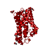
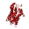
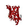
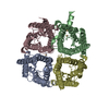
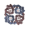
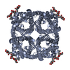
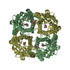
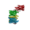
- PDB-3c02: X-ray structure of the aquaglyceroporin from Plasmodium falciparum -

+
Open data


ID or keywords:

Loading...

-
Basic information

Entry
Database: PDB / ID: 3c02
TitleX-ray structure of the aquaglyceroporin from Plasmodium falciparum
ComponentsAquaglyceroporin
KeywordsMEMBRANE PROTEIN / glycerol / water / transport / Structural Genomics / PSI-2 / Protein Structure Initiative / Center for Structures of Membrane Proteins / CSMP / Porin / Transmembrane
Function / homology
Function and homology information


channel activity / membrane => GO:0016020 / identical protein binding
Similarity search - Function
Glycerol uptake facilitator protein / Glycerol uptake facilitator protein. / Major intrinsic protein / Major intrinsic protein / Aquaporin-like / Up-down Bundle / Mainly Alpha
Similarity search - Domain/homology
Biological speciesPlasmodium falciparum (malaria parasite P. falciparum)
MethodX-RAY DIFFRACTION / SYNCHROTRON / MOLECULAR REPLACEMENT / Resolution: 2.05 Å
AuthorsNewby, Z.E. / Stroud, R.M. / Center for Structures of Membrane Proteins (CSMP)
CitationJournal: Nat.Struct.Mol.Biol. / Year: 2008
Title: Crystal structure of the aquaglyceroporin PfAQP from the malarial parasite Plasmodium falciparum.
Authors: Newby, Z.E. / O'Connell, J. / Robles-Colmenares, Y. / Khademi, S. / Miercke, L.J. / Stroud, R.M.
History
DepositionJan 18, 2008Deposition site: RCSB / Processing site: RCSB
Revision 1.0May 27, 2008Provider: repository / Type: Initial release
Revision 1.1Jul 13, 2011Group: Advisory / Version format compliance
Revision 1.2Jul 29, 2020Group: Data collection / Derived calculations / Structure summary
Category: chem_comp / entity ...chem_comp / entity / pdbx_chem_comp_identifier / pdbx_entity_nonpoly / struct_site / struct_site_gen
Item: _chem_comp.mon_nstd_flag / _chem_comp.name ..._chem_comp.mon_nstd_flag / _chem_comp.name / _chem_comp.type / _entity.pdbx_description / _pdbx_entity_nonpoly.name
Description: Carbohydrate remediation / Provider: repository / Type: Remediation
Revision 1.3Aug 30, 2023Group: Data collection / Database references ...Data collection / Database references / Refinement description / Structure summary
Category: chem_comp / chem_comp_atom ...chem_comp / chem_comp_atom / chem_comp_bond / database_2 / pdbx_initial_refinement_model
Item: _chem_comp.pdbx_synonyms / _database_2.pdbx_DOI / _database_2.pdbx_database_accession

-
Structure visualization

Structure viewerMolecule:
MolmilJmol/JSmol

Downloads & links

-
Assembly

Deposited unit
A: Aquaglyceroporin
hetero molecules


Theoretical massNumber of molelcules
Total (without water)29,0727
Polymers28,3191
Non-polymers7536
Water1,00956
1
A: Aquaglyceroporin
hetero molecules

A: Aquaglyceroporin
hetero molecules

A: Aquaglyceroporin
hetero molecules

A: Aquaglyceroporin
hetero molecules


Theoretical massNumber of molelcules
Total (without water)116,28828
Polymers113,2764
Non-polymers3,01124
Water724
TypeNameSymmetry operationNumber
identity operation1_555x,y,z1
crystal symmetry operation2_555-x,-y,z1
crystal symmetry operation3_555-y,x,z1
crystal symmetry operation4_555y,-x,z1
Buried area12670 Å2
MethodPISA
Unit cell
Length a, b, c (Å)92.430, 92.430, 193.168
Angle α, β, γ (deg.)90.00, 90.00, 90.00
Int Tables number97
Space group name H-MI422

-
Components

#1: Protein Aquaglyceroporin


Mass: 28319.070 Da / Num. of mol.: 1
Source method: isolated from a genetically manipulated source
Source: (gene. exp.) Plasmodium falciparum (malaria parasite P. falciparum)
Gene: AQP / Plasmid: pET29b / Production host: Escherichia coli (E. coli) / Strain (production host): C43(DE3) / References: UniProt: Q8WPZ6
#2: Sugar ChemComp-BOG / octyl beta-D-glucopyranoside / Beta-Octylglucoside / octyl beta-D-glucoside / octyl D-glucoside / octyl glucoside


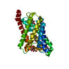
Type: D-saccharide / Mass: 292.369 Da / Num. of mol.: 1
Source method: isolated from a genetically manipulated source
Formula: C14H28O6 / Comment: detergent*YM
IdentifierTypeProgram
b-octylglucosideIUPAC CARBOHYDRATE SYMBOLPDB-CARE 1.0
#3: Chemical
ChemComp-GOL / GLYCEROL / GLYCERIN / PROPANE-1,2,3-TRIOL


Mass: 92.094 Da / Num. of mol.: 5 / Source method: obtained synthetically / Formula: C3H8O3
#4: Water ChemComp-HOH / water


Mass: 18.015 Da / Num. of mol.: 56 / Source method: isolated from a natural source / Formula: H2O

-
Experimental details

-
Experiment

ExperimentMethod: X-RAY DIFFRACTION / Number of used crystals: 1

-
Sample preparation

CrystalDensity Matthews: 3.64 Å3/Da / Density % sol: 66.23 %
Crystal growTemperature: 291 K / Method: vapor diffusion, hanging drop / pH: 5.8
Details: 17% PEG MME 2000, 0.1M Sodium citrate, 15% Glycerol, 0.04M n-octyl-B-D-glucopyranoside, 0.01 Nickel sulfate, 0.01M DTT, pH 5.8, VAPOR DIFFUSION, HANGING DROP, temperature 291K

-
Data collection

DiffractionMean temperature: 100 K
Diffraction sourceSource: SYNCHROTRON / Site: ALS / Beamline: 8.3.1 / Wavelength: 1.11587 Å
DetectorType: ADSC QUANTUM 315 / Detector: CCD / Date: Jul 15, 2007
RadiationMonochromator: Double flat crystal, Si(111) / Protocol: SINGLE WAVELENGTH / Monochromatic (M) / Laue (L): M / Scattering type: x-ray
Radiation wavelengthWavelength: 1.11587 Å / Relative weight: 1
ReflectionResolution: 2.05→46.2 Å / Num. all: 25478 / Num. obs: 24719 / % possible obs: 95.5 % / Observed criterion σ(F): 1 / Observed criterion σ(I): 1 / Redundancy: 15 % / Biso Wilson estimate: 47.9 Å2 / Rmerge(I) obs: 0.084 / Rsym value: 0.06 / Net I/σ(I): 28.8
Reflection shellResolution: 2.05→2.12 Å / Redundancy: 8.1 % / Rmerge(I) obs: 0.478 / Mean I/σ(I) obs: 2.25 / Num. unique all: 1819 / Rsym value: 0.435 / % possible all: 69.9

-
Processing

Software
NameVersionClassification
REFMAC5.3.0037refinement
ADSCQuantumdata collection
HKL-2000data reduction
HKL-2000data scaling
PHASERphasing
RefinementMethod to determine structure: MOLECULAR REPLACEMENT
Starting model: PDB entry 1FX8
Resolution: 2.05→46.2 Å / Cor.coef. Fo:Fc: 0.963 / Cor.coef. Fo:Fc free: 0.961 / SU B: 7.214 / SU ML: 0.094 / TLS residual ADP flag: LIKELY RESIDUAL / Isotropic thermal model: isotropic / Cross valid method: THROUGHOUT / σ(F): 1 / σ(I): 1 / ESU R: 0.134 / ESU R Free: 0.12 / Stereochemistry target values: MAXIMUM LIKELIHOOD / Details: HYDROGENS HAVE BEEN ADDED IN THE RIDING POSITIONS
RfactorNum. reflection% reflectionSelection details
Rfree0.19824 1299 5.1 %RANDOM
Rwork0.18088 ---
all0.184 24719 --
obs0.18175 24179 95.65 %-
Solvent computationIon probe radii: 0.8 Å / Shrinkage radii: 0.8 Å / VDW probe radii: 1.2 Å / Solvent model: MASK
Displacement parametersBiso mean: 37.452 Å2
Baniso -1Baniso -2Baniso -3
1-1.51 Å20 Å20 Å2
2--1.51 Å20 Å2
3----3.02 Å2
Refinement stepCycle: LAST / Resolution: 2.05→46.2 Å
ProteinNucleic acidLigandSolventTotal
Num. atoms1853 0 50 56 1959
Refine LS restraints
Refine-IDTypeDev idealDev ideal targetNumber
X-RAY DIFFRACTIONr_bond_refined_d0.0180.0221981
X-RAY DIFFRACTIONr_bond_other_d
X-RAY DIFFRACTIONr_angle_refined_deg1.6281.9752696
X-RAY DIFFRACTIONr_angle_other_deg
X-RAY DIFFRACTIONr_dihedral_angle_1_deg5.3745253
X-RAY DIFFRACTIONr_dihedral_angle_2_deg35.08323.28670
X-RAY DIFFRACTIONr_dihedral_angle_3_deg13.64615301
X-RAY DIFFRACTIONr_dihedral_angle_4_deg13.794154
X-RAY DIFFRACTIONr_chiral_restr0.1260.2324
X-RAY DIFFRACTIONr_gen_planes_refined0.0070.021436
X-RAY DIFFRACTIONr_gen_planes_other
X-RAY DIFFRACTIONr_nbd_refined0.2240.21089
X-RAY DIFFRACTIONr_nbd_other
X-RAY DIFFRACTIONr_nbtor_refined0.3180.21457
X-RAY DIFFRACTIONr_nbtor_other
X-RAY DIFFRACTIONr_xyhbond_nbd_refined0.1120.296
X-RAY DIFFRACTIONr_xyhbond_nbd_other
X-RAY DIFFRACTIONr_metal_ion_refined
X-RAY DIFFRACTIONr_metal_ion_other
X-RAY DIFFRACTIONr_symmetry_vdw_refined0.190.256
X-RAY DIFFRACTIONr_symmetry_vdw_other
X-RAY DIFFRACTIONr_symmetry_hbond_refined0.2180.23
X-RAY DIFFRACTIONr_symmetry_hbond_other
X-RAY DIFFRACTIONr_symmetry_metal_ion_refined
X-RAY DIFFRACTIONr_symmetry_metal_ion_other
X-RAY DIFFRACTIONr_mcbond_it0.8541.51249
X-RAY DIFFRACTIONr_mcbond_other
X-RAY DIFFRACTIONr_mcangle_it1.32921958
X-RAY DIFFRACTIONr_scbond_it2.3953846
X-RAY DIFFRACTIONr_scangle_it3.3464.5731
X-RAY DIFFRACTIONr_rigid_bond_restr
X-RAY DIFFRACTIONr_sphericity_free
X-RAY DIFFRACTIONr_sphericity_bonded
LS refinement shellResolution: 2.05→2.11 Å / Total num. of bins used: 20
RfactorNum. reflection% reflection
Rfree0.242 67 -
Rwork0.232 1231 -
obs--67.18 %
Refinement TLS params.Method: refined / Origin x: 8.552 Å / Origin y: 19.124 Å / Origin z: 22.738 Å
111213212223313233
T0.2831 Å2-0.0816 Å2-0.0288 Å2-0.1454 Å2-0.1112 Å2--0.0286 Å2
L1.2542 °2-0.1237 °2-0.0528 °2-1.079 °20.0185 °2--1.9651 °2
S-0.0163 Å °-0.3277 Å °0.2916 Å °0.2784 Å °0.0253 Å °-0.1287 Å °-0.519 Å °0.237 Å °-0.0089 Å °

+
About Yorodumi

-
News

-
Feb 9, 2022. New format data for meta-information of EMDB entries

New format data for meta-information of EMDB entries

  • Version 3 of the EMDB header file is now the official format.
  • The previous official version 1.9 will be removed from the archive.

Related info.:EMDB header

External links:wwPDB to switch to version 3 of the EMDB data model

-
Aug 12, 2020. Covid-19 info

Covid-19 info

URL: https://pdbjlvh1.pdbj.org/emnavi/covid19.php

New page: Covid-19 featured information page in EM Navigator.

Related info.:Covid-19 info / Mar 5, 2020. Novel coronavirus structure data

+
Mar 5, 2020. Novel coronavirus structure data

Novel coronavirus structure data

Related info.:Yorodumi Speices / Aug 12, 2020. Covid-19 info

External links:COVID-19 featured content - PDBj / Molecule of the Month (242):Coronavirus Proteases

+
Jan 31, 2019. EMDB accession codes are about to change! (news from PDBe EMDB page)

EMDB accession codes are about to change! (news from PDBe EMDB page)

  • The allocation of 4 digits for EMDB accession codes will soon come to an end. Whilst these codes will remain in use, new EMDB accession codes will include an additional digit and will expand incrementally as the available range of codes is exhausted. The current 4-digit format prefixed with “EMD-” (i.e. EMD-XXXX) will advance to a 5-digit format (i.e. EMD-XXXXX), and so on. It is currently estimated that the 4-digit codes will be depleted around Spring 2019, at which point the 5-digit format will come into force.
  • The EM Navigator/Yorodumi systems omit the EMD- prefix.

Related info.:Q: What is EMD? / ID/Accession-code notation in Yorodumi/EM Navigator

External links:EMDB Accession Codes are Changing Soon! / Contact to PDBj

+
Jul 12, 2017. Major update of PDB

Major update of PDB

  • wwPDB released updated PDB data conforming to the new PDBx/mmCIF dictionary.
  • This is a major update changing the version number from 4 to 5, and with Remediation, in which all the entries are updated.
  • In this update, many items about electron microscopy experimental information are reorganized (e.g. em_software).
  • Now, EM Navigator and Yorodumi are based on the updated data.

External links:wwPDB Remediation / Enriched Model Files Conforming to OneDep Data Standards Now Available in the PDB FTP Archive

-
Yorodumi

Thousand views of thousand structures

  • Yorodumi is a browser for structure data from EMDB, PDB, SASBDB, etc.
  • This page is also the successor to EM Navigator detail page, and also detail information page/front-end page for Omokage search.
  • The word "yorodu" (or yorozu) is an old Japanese word meaning "ten thousand". "mi" (miru) is to see.

Related info.:EMDB / PDB / SASBDB / Comparison of 3 databanks / Yorodumi Search / Aug 31, 2016. New EM Navigator & Yorodumi / Yorodumi Papers / Jmol/JSmol / Function and homology information / Changes in new EM Navigator and Yorodumi

Read more