 Movie
Movie Controller


+ Open data

- Basic information
| Entry | Database: PDB / ID: 7a1h | ||||||
|---|---|---|---|---|---|---|---|
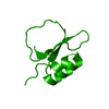
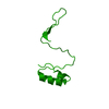
| Title | Crystal structure of wild-type CI2 | ||||||
 Components Components | Subtilisin-chymotrypsin inhibitor-2A | ||||||
 Keywords Keywords | PROTEIN BINDING / Protease inhibitor | ||||||
| Function / homology | Proteinase inhibitor I13, potato inhibitor I / Proteinase inhibitor I13, potato inhibitor I superfamily / Potato inhibitor I family / Potato inhibitor I family signature. / serine-type endopeptidase inhibitor activity / response to wounding / Subtilisin-chymotrypsin inhibitor-2A Function and homology information Function and homology information | ||||||
| Biological species |  | ||||||
| Method |  X-RAY DIFFRACTION / MOLECULAR REPLACEMENT / molecular replacement / Resolution: 1.9 Å X-RAY DIFFRACTION / MOLECULAR REPLACEMENT / molecular replacement / Resolution: 1.9 Å | ||||||
 Authors Authors | Olsen, J.G. / Teilum, K. / Hamborg, L. / Roche, J.V. | ||||||
| Funding support |  Denmark, 1items Denmark, 1items
| ||||||
 Citation Citation | Journal: Commun Biol / Year: 2021 Title: Synergistic stabilization of a double mutant in chymotrypsin inhibitor 2 from a library screen in E. coli. Authors: Hamborg, L. / Granata, D. / Olsen, J.G. / Roche, J.V. / Pedersen, L.E. / Nielsen, A.T. / Lindorff-Larsen, K. / Teilum, K. #1: Journal: Biorxiv / Year: 2020Title: Synergistic stabilization of a double mutant in CI2 from an in-cell library screen Authors: Hamborg, L. / Granata, D. / Olsen, J.G. / Roche, J.V. / Pedersen, L.E. / Nielsen, A.T. / Lindorff-Larsen, K. / Teilum, K. | ||||||
| History |
|
- Structure visualization
| Structure viewer | Molecule: MolmilJmol/JSmol |
|---|
- Downloads & links
-Download
| PDBx/mmCIF format | 7a1h.cif.gz | 28.2 KB | Display | PDBx/mmCIF format |
|---|---|---|---|---|
| PDB format | pdb7a1h.ent.gz | 16.5 KB | Display | PDB format |
| PDBx/mmJSON format | 7a1h.json.gz | Tree view | PDBx/mmJSON format | |
| Others |  Other downloads Other downloads |
-Validation report
| Arichive directory | https://data.pdbj.org/pub/pdb/validation_reports/a1/7a1hftp://data.pdbj.org/pub/pdb/validation_reports/a1/7a1h | HTTPS FTP |
|---|
-Related structure data
| Related structure data | 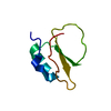 7a3mC  7aokC 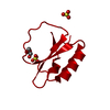 7aonC 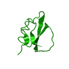 2ci2S S: Starting model for refinement C: citing same article ( |
|---|---|
| Similar structure data |


-Links
 PDBj
PDBj- Assembly
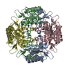
| Deposited unit | 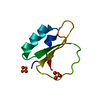
| ||||||||
|---|---|---|---|---|---|---|---|---|---|
| 1 | x 12
| ||||||||
| Unit cell |
|
-Components
| #1: Protein | Mass: 7313.623 Da / Num. of mol.: 1 Source method: isolated from a genetically manipulated source Source: (gene. exp.) Production host:  References: UniProt: P01053 | ||||
|---|---|---|---|---|---|
| #2: Chemical |   Mass: 96.063 Da / Num. of mol.: 2 / Source method: obtained synthetically / Formula: SO4 Mass: 96.063 Da / Num. of mol.: 2 / Source method: obtained synthetically / Formula: SO4#3: Water | ChemComp-HOH / |  Mass: 18.015 Da / Num. of mol.: 30 / Source method: isolated from a natural source / Formula: H2O Mass: 18.015 Da / Num. of mol.: 30 / Source method: isolated from a natural source / Formula: H2OHas ligand of interest | N | |
-Experimental details
-Experiment
| Experiment | Method: X-RAY DIFFRACTION / Number of used crystals: 1 |
|---|
- Sample preparation
| Crystal | Density Matthews: 2.42 Å3/Da / Density % sol: 49.25 % Description: Beautiful three dimensional crystals with clear hexagonal faces. Size of crystals ranging from a few micrometer to app. 1 mm. Become blue when soaked with "Izit". |
|---|---|
| Crystal grow | Temperature: 293 K / Method: vapor diffusion, hanging drop / pH: 8 Details: 40 % (NH4)2SO4, 50 mM Tris-Hcl, pH 8.0. Protein concentration app 75 mg/mL. |
-Data collection
| Diffraction | Mean temperature: 100 K / Serial crystal experiment: N |
|---|---|
| Diffraction source | Source: ROTATING ANODE / Type: Agilent SuperNova / Wavelength: 1.5406 Å |
| Detector | Type: AGILENT ATLAS CCD / Detector: CCD / Date: May 9, 2019 / Details: multilayer |
| Radiation | Protocol: SINGLE WAVELENGTH / Monochromatic (M) / Laue (L): M / Scattering type: x-ray |
| Radiation wavelength | Wavelength: 1.5406 Å / Relative weight: 1 |
| Reflection | Resolution: 1.9→15 Å / Num. obs: 6063 / % possible obs: 99.7 % / Redundancy: 8.7 % / CC1/2: 0.999 / Rpim(I) all: 0.033 / Net I/σ(I): 10.4 |
| Reflection shell | Resolution: 1.9→1.949 Å / Mean I/σ(I) obs: 1.7 / Num. unique obs: 453 / CC1/2: 0.565 / Rpim(I) all: 0.332 / % possible all: 99.8 |
-Phasing
| Phasing | Method: molecular replacement | |||||||||
|---|---|---|---|---|---|---|---|---|---|---|
| Phasing MR | Model details: Phaser MODE: MR_AUTO
|
- Processing
| Software |
| ||||||||||||||||||||||||||||||||||||||||||||||||||||||||||||
|---|---|---|---|---|---|---|---|---|---|---|---|---|---|---|---|---|---|---|---|---|---|---|---|---|---|---|---|---|---|---|---|---|---|---|---|---|---|---|---|---|---|---|---|---|---|---|---|---|---|---|---|---|---|---|---|---|---|---|---|---|---|
| Refinement | Method to determine structure: MOLECULAR REPLACEMENT Starting model: 2CI2 Resolution: 1.9→15 Å / Cor.coef. Fo:Fc: 0.95 / Cor.coef. Fo:Fc free: 0.922 / SU B: 3.733 / SU ML: 0.109 / Cross valid method: THROUGHOUT / σ(F): 0 / ESU R: 0.155 / ESU R Free: 0.152 / Stereochemistry target values: MAXIMUM LIKELIHOOD Details: HYDROGENS HAVE BEEN ADDED IN THE RIDING POSITIONS U VALUES : REFINED INDIVIDUALLY
| ||||||||||||||||||||||||||||||||||||||||||||||||||||||||||||
| Solvent computation | Ion probe radii: 0.8 Å / Shrinkage radii: 0.8 Å / VDW probe radii: 1.2 Å / Solvent model: MASK | ||||||||||||||||||||||||||||||||||||||||||||||||||||||||||||
| Displacement parameters | Biso max: 69.81 Å2 / Biso mean: 23.35 Å2 / Biso min: 10.05 Å2
| ||||||||||||||||||||||||||||||||||||||||||||||||||||||||||||
| Refinement step | Cycle: final / Resolution: 1.9→15 Å
| ||||||||||||||||||||||||||||||||||||||||||||||||||||||||||||
| Refine LS restraints |
| ||||||||||||||||||||||||||||||||||||||||||||||||||||||||||||
| LS refinement shell | Resolution: 1.9→1.949 Å / Rfactor Rfree error: 0 / Total num. of bins used: 20
|
