Movie
Movie Controller

[English] 日本語
 Yorodumi
- PDB-7whp: Pentameric turret of Bombyx mori cytoplasmic polyhedrosis virus a... -
+ Open data

- Basic information
| Entry | Database: PDB / ID: 7whp | |||||||||||||||||||||||||||||||||
|---|---|---|---|---|---|---|---|---|---|---|---|---|---|---|---|---|---|---|---|---|---|---|---|---|---|---|---|---|---|---|---|---|---|---|
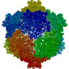
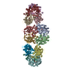
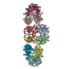
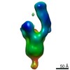
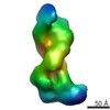
| Title | Pentameric turret of Bombyx mori cytoplasmic polyhedrosis virus after spike detaches. | |||||||||||||||||||||||||||||||||
 Components Components | Structural protein VP3 | |||||||||||||||||||||||||||||||||
 Keywords Keywords | VIRAL PROTEIN / Capping enzyme | |||||||||||||||||||||||||||||||||
| Function / homology |  Function and homology information Function and homology information: / : / : / : / Reovirus VP3 protein, guanylyltransferase (GTase) / Reovirus turret protein, bridge domain / Reovirus VP3 protein, Methyltransferase domain 1 / Reovirus VP3 protein, Methyltransferase domain 2 Similarity search - Domain/homology | |||||||||||||||||||||||||||||||||
| Biological species |   Bombyx mori cypovirus 1 Bombyx mori cypovirus 1 | |||||||||||||||||||||||||||||||||
| Method | ELECTRON MICROSCOPY / single particle reconstruction / cryo EM / Resolution: 3.7 Å | |||||||||||||||||||||||||||||||||
 Authors Authors | Zhang, Y. / Cui, Y. / Sun, J. / Zhou, Z.H. | |||||||||||||||||||||||||||||||||
| Funding support |  China, China,  United States, 10items United States, 10items
| |||||||||||||||||||||||||||||||||
 Citation Citation | Journal: Nat Commun / Year: 2022 Title: Multiple conformations of trimeric spikes visualized on a non-enveloped virus. Authors: Yinong Zhang / Yanxiang Cui / Jingchen Sun / Z Hong Zhou / Abstract: Many viruses utilize trimeric spikes to gain entry into host cells. However, without in situ structures of these trimeric spikes, a full understanding of this dynamic and essential process of viral ...Many viruses utilize trimeric spikes to gain entry into host cells. However, without in situ structures of these trimeric spikes, a full understanding of this dynamic and essential process of viral infections is not possible. Here we present four in situ and one isolated cryoEM structures of the trimeric spike of the cytoplasmic polyhedrosis virus, a member of the non-enveloped Reoviridae family and a virus historically used as a model in the discoveries of RNA transcription and capping. These structures adopt two drastically different conformations, closed spike and opened spike, which respectively represent the penetration-inactive and penetration-active states. Each spike monomer has four domains: N-terminal, body, claw, and C-terminal. From closed to opened state, the RGD motif-containing C-terminal domain is freed to bind integrins, and the claw domain rotates to expose and project its membrane insertion loops into the cellular membrane. Comparison between turret vertices before and after detachment of the trimeric spike shows that the trimeric spike anchors its N-terminal domain in the iris of the pentameric RNA-capping turret. Sensing of cytosolic S-adenosylmethionine (SAM) and adenosine triphosphate (ATP) by the turret triggers a cascade of events: opening of the iris, detachment of the spike, and initiation of endogenous transcription. | |||||||||||||||||||||||||||||||||
| History |
|
- Structure visualization
| Movie |
Movie viewer |
|---|---|
| Structure viewer | Molecule: MolmilJmol/JSmol |
- Downloads & links
-Download
| PDBx/mmCIF format | 7whp.cif.gz | 376.8 KB | Display | PDBx/mmCIF format |
|---|---|---|---|---|
| PDB format | pdb7whp.ent.gz | 304.9 KB | Display | PDB format |
| PDBx/mmJSON format | 7whp.json.gz | Tree view | PDBx/mmJSON format | |
| Others |  Other downloads Other downloads |
-Validation report
| Arichive directory | https://data.pdbj.org/pub/pdb/validation_reports/wh/7whpftp://data.pdbj.org/pub/pdb/validation_reports/wh/7whp | HTTPS FTP |
|---|
-Related structure data
| Related structure data |  32507MC  7whmC  7whnC M: map data used to model this data C: citing same article ( |
|---|---|
| Similar structure data |


-Links
 PDBj
PDBj
- Assembly
| Deposited unit | 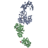
|
|---|---|
| 1 |
|
-Components
| #1: Protein | Mass: 120145.797 Da / Num. of mol.: 2 / Source method: isolated from a natural source / Source: (natural) Bombyx mori cypovirus 1 / References: UniProt: Q914N6#2: Chemical | ChemComp-SAM /   Mass: 398.437 Da / Num. of mol.: 4 / Source method: obtained synthetically / Formula: C15H22N6O5S / Feature type: SUBJECT OF INVESTIGATION Mass: 398.437 Da / Num. of mol.: 4 / Source method: obtained synthetically / Formula: C15H22N6O5S / Feature type: SUBJECT OF INVESTIGATION#3: Chemical |   Mass: 507.181 Da / Num. of mol.: 2 / Source method: obtained synthetically / Formula: C10H16N5O13P3 / Feature type: SUBJECT OF INVESTIGATION / Comment: ATP, energy-carrying molecule*YM Mass: 507.181 Da / Num. of mol.: 2 / Source method: obtained synthetically / Formula: C10H16N5O13P3 / Feature type: SUBJECT OF INVESTIGATION / Comment: ATP, energy-carrying molecule*YMHas ligand of interest | Y | Has protein modification | N | |
|---|
-Experimental details
-Experiment
| Experiment | Method: ELECTRON MICROSCOPY |
|---|---|
| EM experiment | Aggregation state: PARTICLE / 3D reconstruction method: single particle reconstruction |
- Sample preparation
| Component | Name: Bombyx mori cypovirus 1 / Type: VIRUS / Entity ID: #1 / Source: NATURAL | ||||||||||||||||||||||||||||||
|---|---|---|---|---|---|---|---|---|---|---|---|---|---|---|---|---|---|---|---|---|---|---|---|---|---|---|---|---|---|---|---|
| Source (natural) | Organism: Bombyx mori cypovirus 1 | ||||||||||||||||||||||||||||||
| Details of virus | Empty: NO / Enveloped: NO / Isolate: STRAIN / Type: VIRION | ||||||||||||||||||||||||||||||
| Natural host | Organism: Bombyx mori | ||||||||||||||||||||||||||||||
| Buffer solution | pH: 8 | ||||||||||||||||||||||||||||||
| Buffer component |
| ||||||||||||||||||||||||||||||
| Specimen | Embedding applied: NO / Shadowing applied: NO / Staining applied: NO / Vitrification applied: YES | ||||||||||||||||||||||||||||||
| Vitrification | Cryogen name: ETHANE-PROPANE |
- Electron microscopy imaging
| Experimental equipment |  Model: Titan Krios / Image courtesy: FEI Company |
|---|---|
| Microscopy | Model: FEI TITAN KRIOS |
| Electron gun | Electron source:  FIELD EMISSION GUN / Accelerating voltage: 300 kV / Illumination mode: FLOOD BEAM FIELD EMISSION GUN / Accelerating voltage: 300 kV / Illumination mode: FLOOD BEAM |
| Electron lens | Mode: BRIGHT FIELD / Nominal magnification: 130000 X / Nominal defocus max: 2300 nm / Nominal defocus min: 1000 nm |
| Image recording | Electron dose: 40 e/Å2 / Detector mode: SUPER-RESOLUTION / Film or detector model: GATAN K2 QUANTUM (4k x 4k) |
- Processing
| Software | Name: PHENIX / Version: 1.19.2_4158: / Classification: refinement | ||||||||||||||||||||||||
|---|---|---|---|---|---|---|---|---|---|---|---|---|---|---|---|---|---|---|---|---|---|---|---|---|---|
| EM software | Name: PHENIX / Category: model refinement | ||||||||||||||||||||||||
| CTF correction | Type: PHASE FLIPPING AND AMPLITUDE CORRECTION | ||||||||||||||||||||||||
| Symmetry | Point symmetry: C5 (5 fold cyclic) | ||||||||||||||||||||||||
| 3D reconstruction | Resolution: 3.7 Å / Resolution method: FSC 0.143 CUT-OFF / Num. of particles: 3152 / Symmetry type: POINT | ||||||||||||||||||||||||
| Atomic model building | Protocol: FLEXIBLE FIT / Space: REAL | ||||||||||||||||||||||||
| Atomic model building | PDB-ID: 3JB3 Pdb chain-ID: A / Accession code: 3JB3 / Pdb chain residue range: 1-1057 / Source name: PDB / Type: experimental model | ||||||||||||||||||||||||
| Refine LS restraints |
|