 Movie
Movie Controller
 Structure viewers
 About Yorodumi Papers


+Search query
-Structure paper
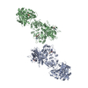
| Title | Multiple conformations of trimeric spikes visualized on a non-enveloped virus. |
|---|---|
| Journal, issue, pages | Nat Commun, Vol. 13, Issue 1, Page 550, Year 2022 |
| Publish date | Jan 27, 2022 |
 Authors Authors | Yinong Zhang / Yanxiang Cui / Jingchen Sun / Z Hong Zhou /   |
| PubMed Abstract | Many viruses utilize trimeric spikes to gain entry into host cells. However, without in situ structures of these trimeric spikes, a full understanding of this dynamic and essential process of viral ...Many viruses utilize trimeric spikes to gain entry into host cells. However, without in situ structures of these trimeric spikes, a full understanding of this dynamic and essential process of viral infections is not possible. Here we present four in situ and one isolated cryoEM structures of the trimeric spike of the cytoplasmic polyhedrosis virus, a member of the non-enveloped Reoviridae family and a virus historically used as a model in the discoveries of RNA transcription and capping. These structures adopt two drastically different conformations, closed spike and opened spike, which respectively represent the penetration-inactive and penetration-active states. Each spike monomer has four domains: N-terminal, body, claw, and C-terminal. From closed to opened state, the RGD motif-containing C-terminal domain is freed to bind integrins, and the claw domain rotates to expose and project its membrane insertion loops into the cellular membrane. Comparison between turret vertices before and after detachment of the trimeric spike shows that the trimeric spike anchors its N-terminal domain in the iris of the pentameric RNA-capping turret. Sensing of cytosolic S-adenosylmethionine (SAM) and adenosine triphosphate (ATP) by the turret triggers a cascade of events: opening of the iris, detachment of the spike, and initiation of endogenous transcription. |
 External links External links | Nat Commun / PubMed:35087065 / PubMed Central |
| Methods | EM (single particle) |
| Resolution | 2.7 - 4.1 Å |
| Structure data | EMDB-32504, PDB-7whm: EMDB-32505, PDB-7whn:  EMDB-32506: EMDB-32507, PDB-7whp:  EMDB-32508: |
| Chemicals |  ChemComp-SAM:  ChemComp-ATP: |
| Source |
|
 Keywords Keywords | VIRAL PROTEIN / Cell attachment / Membrane penetration / Capping enzyme |


 bombyx mori cypovirus 1