 Movie
Movie Controller

[English] 日本語
 Yorodumi
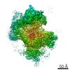
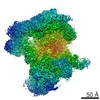
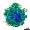
- EMDB-32506: 5-fold vertex of Bombyx mori cytoplasmic polyhedrosis virus forme... -
+ Open data

- Basic information
| Entry | Database: EMDB / ID: EMD-32506 | |||||||||||||||||||||||||||||||||
|---|---|---|---|---|---|---|---|---|---|---|---|---|---|---|---|---|---|---|---|---|---|---|---|---|---|---|---|---|---|---|---|---|---|---|
| Title | 5-fold vertex of Bombyx mori cytoplasmic polyhedrosis virus formed by trimeric spike and pentameric turret | |||||||||||||||||||||||||||||||||
 Map data Map data | ||||||||||||||||||||||||||||||||||
 Sample Sample |
| |||||||||||||||||||||||||||||||||
 Keywords Keywords | Complex / Cell attachment / Membrane penetration / Capping enzyme / VIRAL PROTEIN | |||||||||||||||||||||||||||||||||
| Function / homology |  Function and homology information Function and homology information | |||||||||||||||||||||||||||||||||
| Biological species |   Bombyx mori cypovirus 1 Bombyx mori cypovirus 1 | |||||||||||||||||||||||||||||||||
| Method | single particle reconstruction / cryo EM / Resolution: 4.1 Å | |||||||||||||||||||||||||||||||||
 Authors Authors | Zhang Y / Cui Y / Sun J / Zhou ZH | |||||||||||||||||||||||||||||||||
| Funding support |  China, 10 items China, 10 items
| |||||||||||||||||||||||||||||||||
 Citation Citation | Journal: Nat Commun / Year: 2022 Title: Multiple conformations of trimeric spikes visualized on a non-enveloped virus. Authors: Yinong Zhang / Yanxiang Cui / Jingchen Sun / Z Hong Zhou /  Abstract: Many viruses utilize trimeric spikes to gain entry into host cells. However, without in situ structures of these trimeric spikes, a full understanding of this dynamic and essential process of viral ...Many viruses utilize trimeric spikes to gain entry into host cells. However, without in situ structures of these trimeric spikes, a full understanding of this dynamic and essential process of viral infections is not possible. Here we present four in situ and one isolated cryoEM structures of the trimeric spike of the cytoplasmic polyhedrosis virus, a member of the non-enveloped Reoviridae family and a virus historically used as a model in the discoveries of RNA transcription and capping. These structures adopt two drastically different conformations, closed spike and opened spike, which respectively represent the penetration-inactive and penetration-active states. Each spike monomer has four domains: N-terminal, body, claw, and C-terminal. From closed to opened state, the RGD motif-containing C-terminal domain is freed to bind integrins, and the claw domain rotates to expose and project its membrane insertion loops into the cellular membrane. Comparison between turret vertices before and after detachment of the trimeric spike shows that the trimeric spike anchors its N-terminal domain in the iris of the pentameric RNA-capping turret. Sensing of cytosolic S-adenosylmethionine (SAM) and adenosine triphosphate (ATP) by the turret triggers a cascade of events: opening of the iris, detachment of the spike, and initiation of endogenous transcription. | |||||||||||||||||||||||||||||||||
| History |
|
- Structure visualization
| Movie |
Movie viewer |
|---|---|
| Structure viewer | EM map: SurfViewMolmilJmol/JSmol |
| Supplemental images |

- Downloads & links
-EMDB archive
| Map data | emd_32506.map.gz | 98.6 MB | EMDB map data format | |
|---|---|---|---|---|
| Header (meta data) | emd-32506-v30.xmlemd-32506.xml | 14.8 KB 14.8 KB | Display Display | EMDB header |
| Images | 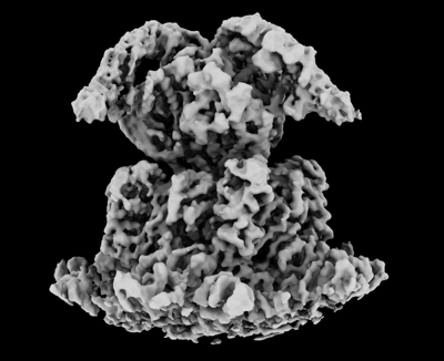 emd_32506.png emd_32506.png | 84.5 KB | ||
| Filedesc metadata | emd-32506.cif.gz | 6.2 KB | ||
| Archive directory |  http://ftp.pdbj.org/pub/emdb/structures/EMD-32506ftp://ftp.pdbj.org/pub/emdb/structures/EMD-32506 http://ftp.pdbj.org/pub/emdb/structures/EMD-32506ftp://ftp.pdbj.org/pub/emdb/structures/EMD-32506 | HTTPS FTP |
-Related structure data


-Links
| EMDB pages | EMDB (EBI/PDBe) / EMDataResource |
|---|
-Map
| File | Download / File: emd_32506.map.gz / Format: CCP4 / Size: 125 MB / Type: IMAGE STORED AS FLOATING POINT NUMBER (4 BYTES) | ||||||||||||||||||||||||||||||||||||||||||||||||||||||||||||||||||||
|---|---|---|---|---|---|---|---|---|---|---|---|---|---|---|---|---|---|---|---|---|---|---|---|---|---|---|---|---|---|---|---|---|---|---|---|---|---|---|---|---|---|---|---|---|---|---|---|---|---|---|---|---|---|---|---|---|---|---|---|---|---|---|---|---|---|---|---|---|---|
| Projections & slices | Image control
Images are generated by Spider. | ||||||||||||||||||||||||||||||||||||||||||||||||||||||||||||||||||||
| Voxel size | X=Y=Z: 1.062 Å | ||||||||||||||||||||||||||||||||||||||||||||||||||||||||||||||||||||
| Density |
| ||||||||||||||||||||||||||||||||||||||||||||||||||||||||||||||||||||
| Symmetry | Space group: 1 | ||||||||||||||||||||||||||||||||||||||||||||||||||||||||||||||||||||
| Details | EMDB XML:
CCP4 map header:
| ||||||||||||||||||||||||||||||||||||||||||||||||||||||||||||||||||||
 Z (Sec.)
 Y (Row.)
 X (Col.)


-Supplemental data
- Sample components
-Entire : Bombyx mori cypovirus 1
| Entire | Name: Bombyx mori cypovirus 1 |
|---|---|
| Components |
|
-Supramolecule #1: Bombyx mori cypovirus 1
| Supramolecule | Name: Bombyx mori cypovirus 1 / type: virus / ID: 1 / Parent: 0 / Macromolecule list: all / NCBI-ID: 110829 / Sci species name: Bombyx mori cypovirus 1 / Virus type: VIRION / Virus isolate: STRAIN / Virus enveloped: No / Virus empty: No |
|---|---|
| Host (natural) | Organism:  |
-Macromolecule #1: Spike protein encoded by S3 gene of Bombyx mori cytoplasmic polyh...
| Macromolecule | Name: Spike protein encoded by S3 gene of Bombyx mori cytoplasmic polyhedrosis virus. type: protein_or_peptide / ID: 1 / Enantiomer: LEVO |
|---|---|
| Source (natural) | Organism: Bombyx mori cypovirus 1 |
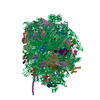
| Sequence | String: MEINRAEIRR EITRYTGLIE QQTQLNISDN DENILKTLIA DYNLRMRRDA LLGELARLDE LRDISQVKGV EYKVTIPLL PVISTLNQHE FEITQANIET DFIADNVTFV TSFVPADLDL EQTIQRVFFR TTATTPHFQS F NLVIEILN YDQDSGDVEL HVKIMIVRPN ...String: MEINRAEIRR EITRYTGLIE QQTQLNISDN DENILKTLIA DYNLRMRRDA LLGELARLDE LRDISQVKGV EYKVTIPLL PVISTLNQHE FEITQANIET DFIADNVTFV TSFVPADLDL EQTIQRVFFR TTATTPHFQS F NLVIEILN YDQDSGDVEL HVKIMIVRPN SDVVNYDYTW IGKDYERISV CYNLISHLQR IDGPHGRDDE AE MPIYRII RRDSGSIPSY ASGEHLYVIS SHLHVDEIVR RREHKSISVD VTQLSLILPI IRTFNPVDLR EVR IEDITP GIEFTINMEV STYLAESSGS HVDMQRAIMN HADKIVGNYT GQQWNVQSNM LSEVRTQMLE EEDE EARQR GDYTTSTLVQ TMAQVSDLFS STILYRRAEA RLDNTVGAFE LLRPVLSIPS EYVHNGRVGP ITNIP ANAS IVTSSSSGAG QVRNIFKPIG DQTINESHFA NVFSNDEYAI YLRFSYRQAP VQSETVYLQQ NLPSMR IVS PSSVSTTVST AVIGGNTIHI NCPIRPHRED RLVSGGVQVP RQSTAVEIRV QEILIGYRQA TTFPIDT EG RLSLELMYGL ESRSAVGNTM SPVRFVTVND GEFFGLTCPI DLTLSTVVDP SSYLSDGVIL VATAFEDL R GYAWVATLGG DWPRTYNSSM RAFNVLTGGD INLSTEYGSE MTYTFKVELP IVYMFNNMTV ISNNVPRVP VLGVTYASIY QDSRTELEAR RFLQTLVFRI HGNWSARIPY TPGNLPTRNT ANQHQDIQQV INDSISQELG RLSDELLNM KNRLDHLERQ FEMFIQSQES EWWEILLNVV MDTVLGYFST FAGNALKSAQ QAISKAVGYT R RVLMTVTK TMRNGPIFTR LLGAKNLSGQ ALASLETLVE SVLRSINVKK SRFMSGAEPL YKNNKVAQHI DN TEKMNMM MDFSFANRNN RQNITADTLS RMHTQNAHGT SDTVLPAMRV YYRPLGFLDK RVGEALHKGI TRP EALKKQ LRSDVANVGT RAPSHAFMTY TDVLYEDAGS YIVSKRYLGI GELNRFGRTT SDKNADIGGV NIKY RVNKI TADGKYIIDR LSHTESGYTA ADVDRLYRSL FGKQGDGLST EQKWMDISRG VDAKIISADM VSEEF LSSK YTGQMIDELI NSPPQFNYSL IYRNCQDFVL DVLRVAQGFS PSNKWDVSTA ARMQQRRVIS LMDDLM SES ETFARSAHSN HSLLQQIRRS YVKARKRGDL HTVKALQLRL KGFFQI |
-Macromolecule #2: Turret protein encoded by S4 gene of Bombyx mori cytoplasmic poly...
| Macromolecule | Name: Turret protein encoded by S4 gene of Bombyx mori cytoplasmic polyhedrosis virus type: protein_or_peptide / ID: 2 / Enantiomer: LEVO |
|---|---|
| Source (natural) | Organism: Bombyx mori cypovirus 1 |
| Sequence | String: MWHYTSINND TRVALDPKPN QIRTITKPNT VPQLGTDYLY TFNSQRRSHT LRLLGPFQYF NFSETDRGHP LFRLPLKYPS KAIPADELI DNLHSWMRSV HLLHVRSEDN TLRYNWMLGV YARSTNYTTP VGQLVVNAPA ILNYSNPQDA FNSVFVALGI D YIDIPITN ...String: MWHYTSINND TRVALDPKPN QIRTITKPNT VPQLGTDYLY TFNSQRRSHT LRLLGPFQYF NFSETDRGHP LFRLPLKYPS KAIPADELI DNLHSWMRSV HLLHVRSEDN TLRYNWMLGV YARSTNYTTP VGQLVVNAPA ILNYSNPQDA FNSVFVALGI D YIDIPITN SNIFDDSSTP YNVRIWHAPT MTEVNHILAL MRKSTLVSTH SSWHWNVLHT FHYRSESDMI DHFAAKILED WR QKEKLDK GALVEADRVI QRLIPLSSST YVQRLAAIGA LYPNEFTENV LDLSRLSTAL LQLSDTYYQH ANDQLRRLYR RMY NDSRTL YMTQRHQELL LAQITADPNI LLYPYTYIFT TIPTSMNYIS NTGQGRIKHS LTVTGATEHD TVADIVLGQT GEDV ITISM VEPMSIAVED MYGYVLDTPT RDIWPADEQI EQKGDAVALY DTKTSRALGM FNNTVRIDDL LSPLLSLVYR TYIKG DTMT MTQGSLDHLT LCAAVDSDIT FVGNRMIAPL PEGYIPKPMH RNNSTMKMLS LYVALKKLEN FATNSYLMAP DTSIIL LGA EREPAVNILR RFNRNVSNVR IIGMGDRAVE PNIRVRVPFP IDKNISADFI ICDINSYEDQ SFESMFSETI SVVTTCA SA ATRALVKINH PSEYMINSVI ERLSQLGGVF YHTALLKTAS QNPYSYETYI YITPIAAAVR FPFYSNSAMI NRYMTAVA D DEMPIIPSIH TVIKGHSNTY SPGLFCGCVD VQSAPLALSQ LKSYCSEATT WRVDSDDNLV NIIARIDPAR IALEFRTRS NTSAYHEYQR YVPNGLGFKV RKTREFRYMH REVTFIHKLM MYALIREQIS LTENMTQVVS IGGRNLADIS VVPLNMKYVV IDPATRIET LTQEKKNIEV QSRPFQFDAA NMDLENNSIY LFIAVIMNEP NGAATPARMQ MDKIRNVATA MLTRTNCVAY I SFYEAGII TRLDQSTAHK TIRVEEGRLK VANYVPVDTL VEADVTLMLR DIGITHEIIR PSTPELIDAC SNYGIRLGST GG AVLDVFN HYSPVIKLVR S |
-Experimental details
-Structure determination
| Method | cryo EM |
|---|---|
 Processing Processing | single particle reconstruction |
| Aggregation state | particle |
-Sample preparation
| Buffer | pH: 8 Component:
| ||||||||||||
|---|---|---|---|---|---|---|---|---|---|---|---|---|---|
| Vitrification | Cryogen name: ETHANE-PROPANE |
- Electron microscopy
| Microscope | FEI TITAN KRIOS |
|---|---|
| Image recording | Film or detector model: GATAN K2 QUANTUM (4k x 4k) / Detector mode: SUPER-RESOLUTION / Average electron dose: 40.0 e/Å2 |
| Electron beam | Acceleration voltage: 300 kV / Electron source:  FIELD EMISSION GUN FIELD EMISSION GUN |
| Electron optics | Illumination mode: FLOOD BEAM / Imaging mode: BRIGHT FIELD / Nominal defocus max: 2.3000000000000003 µm / Nominal defocus min: 1.0 µm / Nominal magnification: 130000 |
| Experimental equipment |  Model: Titan Krios / Image courtesy: FEI Company |
-Image processing
| Startup model | Type of model: NONE |
|---|---|
| Final reconstruction | Applied symmetry - Point group: C1 (asymmetric) / Resolution.type: BY AUTHOR / Resolution: 4.1 Å / Resolution method: FSC 0.143 CUT-OFF / Number images used: 69955 |
| Initial angle assignment | Type: ANGULAR RECONSTITUTION |
| Final angle assignment | Type: ANGULAR RECONSTITUTION |
