+ Open data
- Basic information
| Entry | Database: EMDB / ID: EMD-32505 | |||||||||||||||||||||||||||||||||
|---|---|---|---|---|---|---|---|---|---|---|---|---|---|---|---|---|---|---|---|---|---|---|---|---|---|---|---|---|---|---|---|---|---|---|
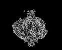
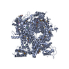
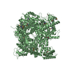
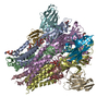
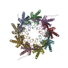
| Title | Opened spike of Bombyx mori cytoplasmic polyhedrosis virus | |||||||||||||||||||||||||||||||||
 Map data Map data | ||||||||||||||||||||||||||||||||||
 Sample Sample |
| |||||||||||||||||||||||||||||||||
 Keywords Keywords | Cell attachment / Membrane penetration / VIRAL PROTEIN | |||||||||||||||||||||||||||||||||
| Function / homology | PPPDE peptidase domain / PPPDE domain profile. / peptidase activity / PPPDE domain-containing protein Function and homology information Function and homology information | |||||||||||||||||||||||||||||||||
| Biological species |   Bombyx mori cypovirus 1 Bombyx mori cypovirus 1 | |||||||||||||||||||||||||||||||||
| Method | single particle reconstruction / cryo EM / Resolution: 3.3 Å | |||||||||||||||||||||||||||||||||
 Authors Authors | Zhang Y / Cui Y | |||||||||||||||||||||||||||||||||
| Funding support |  China, China,  United States, 10 items United States, 10 items
| |||||||||||||||||||||||||||||||||
 Citation Citation |  Journal: Nat Commun / Year: 2022 Journal: Nat Commun / Year: 2022Title: Multiple conformations of trimeric spikes visualized on a non-enveloped virus. Authors: Yinong Zhang / Yanxiang Cui / Jingchen Sun / Z Hong Zhou /   Abstract: Many viruses utilize trimeric spikes to gain entry into host cells. However, without in situ structures of these trimeric spikes, a full understanding of this dynamic and essential process of viral ...Many viruses utilize trimeric spikes to gain entry into host cells. However, without in situ structures of these trimeric spikes, a full understanding of this dynamic and essential process of viral infections is not possible. Here we present four in situ and one isolated cryoEM structures of the trimeric spike of the cytoplasmic polyhedrosis virus, a member of the non-enveloped Reoviridae family and a virus historically used as a model in the discoveries of RNA transcription and capping. These structures adopt two drastically different conformations, closed spike and opened spike, which respectively represent the penetration-inactive and penetration-active states. Each spike monomer has four domains: N-terminal, body, claw, and C-terminal. From closed to opened state, the RGD motif-containing C-terminal domain is freed to bind integrins, and the claw domain rotates to expose and project its membrane insertion loops into the cellular membrane. Comparison between turret vertices before and after detachment of the trimeric spike shows that the trimeric spike anchors its N-terminal domain in the iris of the pentameric RNA-capping turret. Sensing of cytosolic S-adenosylmethionine (SAM) and adenosine triphosphate (ATP) by the turret triggers a cascade of events: opening of the iris, detachment of the spike, and initiation of endogenous transcription. | |||||||||||||||||||||||||||||||||
| History |
|
- Structure visualization
| Movie |
 Movie viewer Movie viewer |
|---|---|
| Structure viewer | EM map:  SurfView SurfView Molmil Molmil Jmol/JSmol Jmol/JSmol |
| Supplemental images |
- Downloads & links
-EMDB archive
| Map data |  emd_32505.map.gz emd_32505.map.gz | 117.2 MB |  EMDB map data format EMDB map data format | |
|---|---|---|---|---|
| Header (meta data) |  emd-32505-v30.xml emd-32505-v30.xml emd-32505.xml emd-32505.xml | 16.4 KB 16.4 KB | Display Display |  EMDB header EMDB header |
| Images |  emd_32505.png emd_32505.png | 59 KB | ||
| Filedesc metadata |  emd-32505.cif.gz emd-32505.cif.gz | 6.6 KB | ||
| Archive directory |  http://ftp.pdbj.org/pub/emdb/structures/EMD-32505 http://ftp.pdbj.org/pub/emdb/structures/EMD-32505 ftp://ftp.pdbj.org/pub/emdb/structures/EMD-32505 ftp://ftp.pdbj.org/pub/emdb/structures/EMD-32505 | HTTPS FTP |
-Validation report
| Summary document |  emd_32505_validation.pdf.gz emd_32505_validation.pdf.gz | 661.8 KB | Display |  EMDB validaton report EMDB validaton report |
|---|---|---|---|---|
| Full document |  emd_32505_full_validation.pdf.gz emd_32505_full_validation.pdf.gz | 661.4 KB | Display | |
| Data in XML |  emd_32505_validation.xml.gz emd_32505_validation.xml.gz | 6.5 KB | Display | |
| Data in CIF |  emd_32505_validation.cif.gz emd_32505_validation.cif.gz | 7.6 KB | Display | |
| Arichive directory |  https://ftp.pdbj.org/pub/emdb/validation_reports/EMD-32505 https://ftp.pdbj.org/pub/emdb/validation_reports/EMD-32505 ftp://ftp.pdbj.org/pub/emdb/validation_reports/EMD-32505 ftp://ftp.pdbj.org/pub/emdb/validation_reports/EMD-32505 | HTTPS FTP |
-Related structure data
| Related structure data |  7whnMC  7whmC  7whpC M: atomic model generated by this map C: citing same article ( |
|---|---|
| Similar structure data |
- Links
Links
| EMDB pages |  EMDB (EBI/PDBe) / EMDB (EBI/PDBe) /  EMDataResource EMDataResource |
|---|
- Map
Map
| File |  Download / File: emd_32505.map.gz / Format: CCP4 / Size: 125 MB / Type: IMAGE STORED AS FLOATING POINT NUMBER (4 BYTES) Download / File: emd_32505.map.gz / Format: CCP4 / Size: 125 MB / Type: IMAGE STORED AS FLOATING POINT NUMBER (4 BYTES) | ||||||||||||||||||||||||||||||||||||||||||||||||||||||||||||||||||||
|---|---|---|---|---|---|---|---|---|---|---|---|---|---|---|---|---|---|---|---|---|---|---|---|---|---|---|---|---|---|---|---|---|---|---|---|---|---|---|---|---|---|---|---|---|---|---|---|---|---|---|---|---|---|---|---|---|---|---|---|---|---|---|---|---|---|---|---|---|---|
| Projections & slices | Image control
Images are generated by Spider. | ||||||||||||||||||||||||||||||||||||||||||||||||||||||||||||||||||||
| Voxel size | X=Y=Z: 1.062 Å | ||||||||||||||||||||||||||||||||||||||||||||||||||||||||||||||||||||
| Density |
| ||||||||||||||||||||||||||||||||||||||||||||||||||||||||||||||||||||
| Symmetry | Space group: 1 | ||||||||||||||||||||||||||||||||||||||||||||||||||||||||||||||||||||
| Details | EMDB XML:
CCP4 map header:
| ||||||||||||||||||||||||||||||||||||||||||||||||||||||||||||||||||||
-Supplemental data
- Sample components
-Entire : Bombyx mori cypovirus 1
| Entire | Name:   Bombyx mori cypovirus 1 Bombyx mori cypovirus 1 |
|---|---|
| Components |
|
-Supramolecule #1: Bombyx mori cypovirus 1
| Supramolecule | Name: Bombyx mori cypovirus 1 / type: virus / ID: 1 / Parent: 0 / Macromolecule list: all / NCBI-ID: 110829 / Sci species name: Bombyx mori cypovirus 1 / Virus type: VIRION / Virus isolate: STRAIN / Virus enveloped: No / Virus empty: No |
|---|---|
| Host (natural) | Organism:  |
-Macromolecule #1: PPPDE domain-containing protein
| Macromolecule | Name: PPPDE domain-containing protein / type: protein_or_peptide / ID: 1 / Number of copies: 1 / Enantiomer: LEVO |
|---|---|
| Source (natural) | Organism:   Bombyx mori cypovirus 1 Bombyx mori cypovirus 1 |
| Molecular weight | Theoretical: 140.37025 KDa |
| Sequence | String: MEINRAEIRR EITRYTGLIE QQTQLNISDN DENILKTLIA DYNLRMRRDA LLGELARLDE LRDISQVKGV EYKVTIPLLP VISTLNQHE FEITQANIET DFIADNVTFV TSFVPADLDL EQTIQRVFFR TTATTPHFQS FNLVIEILNY DQDSGDVELH V KIMIVRPN ...String: MEINRAEIRR EITRYTGLIE QQTQLNISDN DENILKTLIA DYNLRMRRDA LLGELARLDE LRDISQVKGV EYKVTIPLLP VISTLNQHE FEITQANIET DFIADNVTFV TSFVPADLDL EQTIQRVFFR TTATTPHFQS FNLVIEILNY DQDSGDVELH V KIMIVRPN SDVVNYDYTW IGKDYERISV CYNLISHLQR IDGPHGRDDE AEMPIYRIIR RDSGSIPSYA SGEHLYVISS HL HVDEIVR RREHKSISVD VTQLSLILPI IRTFNPVDLR EVRIEDITPG IEFTINMEVS TYLAESSGSH VDMQRAIMNH ADK IVGNYT GQQWNVQSNM LSEVRTQMLE EEDEEARQRG DYTTSTLVQT MAQVSDLFSS TILYRRAEAR LDNTVGAFEL LRPV LSIPS EYVHNGRVGP ITNIPANASI VTSSSSGAGQ VRNIFKPIGD QTINESHFAN VFSNDEYAIY LRFSYRQAPV QSETV YLQQ NLPSMRIVSP SSVSTTVSTA VIGGNTIHIN CPIRPHREDR LVSGGVQVPR QSTAVEIRVQ EILIGYRQAT TFPIDT EGR LSLELMYGLE SRSAVGNTMS PVRFVTVNDG EFFGLTCPID LTLSTVVDPS SYLSDGVILV ATAFEDLRGY AWVATLG GD WPRTYNSSMR AFNVLTGGDI NLSTEYGSEM TYTFKVELPI VYMFNNMTVI SNNVPRVPVL GVTYASIYQD SRTELEAR R FLQTLVFRIH GNWSARIPYT PGNLPTRNTA NQHQDIQQVI NDSISQELGR LSDELLNMKN RLDHLERQFE MFIQSQESE WWEILLNVVM DTVLGYFSTF AGNALKSAQQ AISKAVGYTR RVLMTVTKTM RNGPIFTRLL GAKNLSGQAL ASLETLVESV LRSINVKKS RFMSGAEPLY KNNKVAQHID NTEKMNMMMD FSFANRNNRQ NITADTLSRM HTQNAHGTSD TVLPAMRVYY R PLGFLDKR VGEALHKGIT RPEALKKQLR SDVANVGTRA PSHAFMTYTD VLYEDAGSYI VSKRYLGIGE LNRFGRTTSD KN ADIGGVN IKYRVNKITA DGKYIIDRLS HTESGYTAAD VDRLYRSLFG KQGDGLSTEQ KWMDISRGVD AKIISADMVS EEF LSSKYT GQMIDELINS PPQFNYSLIY RNCQDFVLDV LRVAQGFSPS NKWDVSTAAR MQQRRVISLM DDLMSESETF ARSA HSNHS LLQQIRRSYV KARKRGDLHT VKALQLRLKG FFQI UniProtKB: PPPDE domain-containing protein |
-Experimental details
-Structure determination
| Method | cryo EM |
|---|---|
 Processing Processing | single particle reconstruction |
| Aggregation state | particle |
- Sample preparation
| Buffer | pH: 8 Component:
| ||||||||||||
|---|---|---|---|---|---|---|---|---|---|---|---|---|---|
| Vitrification | Cryogen name: ETHANE-PROPANE |
- Electron microscopy
| Microscope | FEI TITAN KRIOS |
|---|---|
| Image recording | Film or detector model: GATAN K2 QUANTUM (4k x 4k) / Detector mode: SUPER-RESOLUTION / Average electron dose: 40.0 e/Å2 |
| Electron beam | Acceleration voltage: 300 kV / Electron source:  FIELD EMISSION GUN FIELD EMISSION GUN |
| Electron optics | Illumination mode: FLOOD BEAM / Imaging mode: BRIGHT FIELD / Nominal defocus max: 2.3000000000000003 µm / Nominal defocus min: 1.0 µm / Nominal magnification: 130000 |
| Experimental equipment |  Model: Titan Krios / Image courtesy: FEI Company |
+ Image processing
-Atomic model buiding 1
| Refinement | Space: REAL / Protocol: AB INITIO MODEL |
|---|---|
| Output model |  PDB-7whn: |
 Movie
Movie Controller





 Z (Sec.)
 Y (Row.)
 X (Col.)


