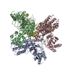
 Movie
Movie Controller


+ Open data

- Basic information
| Entry | Database: PDB / ID: 1p4e | ||||||
|---|---|---|---|---|---|---|---|
| Title | Flpe W330F mutant-DNA Holliday Junction Complex | ||||||
 Components Components |
| ||||||
 Keywords Keywords | DNA BINDING PROTEIN/RECOMBINATION/DNA / Flp / Holliday junction / site-specific recombination / W330 / Flpe / DNA BINDING PROTEIN-RECOMBINATION-DNA COMPLEX | ||||||
| Function / homology |  Function and homology information Function and homology informationplasmid recombination / site-specific recombinase activity / DNA binding, bending / single-stranded DNA binding / double-stranded DNA binding Similarity search - Function | ||||||
| Biological species |  | ||||||
| Method |  X-RAY DIFFRACTION / SYNCHROTRON / FOURIER SYNTHESIS / Resolution: 2.7 Å X-RAY DIFFRACTION / SYNCHROTRON / FOURIER SYNTHESIS / Resolution: 2.7 Å | ||||||
 Authors Authors | Rice, P.A. / Chen, Y. | ||||||
 Citation Citation | Journal: J.Biol.Chem. / Year: 2003 Title: The role of the conserved Trp330 in Flp-mediated recombination. Functional and structural analysis Authors: Rice, P.A. / Chen, Y. | ||||||
| History |
| ||||||
| Remark 999 | The sequence in the database is that of Flp. For this experiment, it is Flpe, a mutant of Flp. ... The sequence in the database is that of Flp. For this experiment, it is Flpe, a mutant of Flp. Flpe differs from Flp with four point mutations at 2, 33, 108 and 294. |
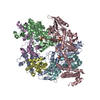
- Structure visualization
| Structure viewer | Molecule: MolmilJmol/JSmol |
|---|
- Downloads & links
-Download
| PDBx/mmCIF format | 1p4e.cif.gz | 406 KB | Display | PDBx/mmCIF format |
|---|---|---|---|---|
| PDB format | pdb1p4e.ent.gz | 324.4 KB | Display | PDB format |
| PDBx/mmJSON format | 1p4e.json.gz | Tree view | PDBx/mmJSON format | |
| Others |  Other downloads Other downloads |
-Validation report
| Arichive directory | https://data.pdbj.org/pub/pdb/validation_reports/p4/1p4eftp://data.pdbj.org/pub/pdb/validation_reports/p4/1p4e | HTTPS FTP |
|---|
-Related structure data
| Related structure data |  1m6xS S: Starting model for refinement |
|---|---|
| Similar structure data |


-Links
 PDBj
PDBj


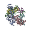
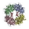
- Assembly
| Deposited unit | 
| ||||||||
|---|---|---|---|---|---|---|---|---|---|
| 1 |
| ||||||||
| Unit cell |
|
-Components
-DNA chain , 3 types, 6 molecules EFIJGH
| #1: DNA chain | Mass: 3916.571 Da / Num. of mol.: 2 / Source method: obtained synthetically #2: DNA chain | Mass: 6165.042 Da / Num. of mol.: 2 / Source method: obtained synthetically #3: DNA chain | Mass: 10126.570 Da / Num. of mol.: 2 / Source method: obtained synthetically Details: 2PO represents 3'-phosphate which attached to C13 of chain H |
|---|
-Recombinase FLP ... , 2 types, 4 molecules ABCD
| #4: Protein | Mass: 49383.395 Da / Num. of mol.: 2 / Fragment: Flpe / Mutation: W330F Source method: isolated from a genetically manipulated source Details: C-terminal (His)6 tag via a glycine linker Source: (gene. exp.) Gene: FLP1 / Plasmid: pET24d / Production host:  #5: Protein | Mass: 49463.375 Da / Num. of mol.: 2 / Fragment: Flpe / Mutation: W330F Source method: isolated from a genetically manipulated source Details: Posttranslational modification of TYR343 to PTR. This modification is a result of covalent binding to DNA. Source: (gene. exp.) Gene: FLP1 / Plasmid: pET24d / Production host: |
|---|
-Non-polymers , 2 types, 267 molecules 

| #6: Chemical | ChemComp-2PO /  Mass: 79.980 Da / Num. of mol.: 1 / Source method: obtained synthetically / Formula: HO3P / Details: 3'-phosphate Mass: 79.980 Da / Num. of mol.: 1 / Source method: obtained synthetically / Formula: HO3P / Details: 3'-phosphate |
|---|---|
| #7: Water | ChemComp-HOH / Mass: 18.015 Da / Num. of mol.: 266 / Source method: isolated from a natural source / Formula: H2O |
-Details
| Has protein modification | Y |
|---|
-Experimental details
-Experiment
| Experiment | Method: X-RAY DIFFRACTION / Number of used crystals: 1 |
|---|
- Sample preparation
| Crystal | Density Matthews: 2.91 Å3/Da / Density % sol: 57.44 % | ||||||||||||||||||||||||||||||||||||||||||||||||||||||||||||||||||||||||||||||||||||||||||||||||||
|---|---|---|---|---|---|---|---|---|---|---|---|---|---|---|---|---|---|---|---|---|---|---|---|---|---|---|---|---|---|---|---|---|---|---|---|---|---|---|---|---|---|---|---|---|---|---|---|---|---|---|---|---|---|---|---|---|---|---|---|---|---|---|---|---|---|---|---|---|---|---|---|---|---|---|---|---|---|---|---|---|---|---|---|---|---|---|---|---|---|---|---|---|---|---|---|---|---|---|---|
| Crystal grow | Temperature: 277 K / Method: vapor diffusion, hanging drop / pH: 7 Details: PEG 5KMME, Hepes, xylitol, sodium chloride, calcium chloride, pH 7.0, VAPOR DIFFUSION, HANGING DROP, temperature 277K | ||||||||||||||||||||||||||||||||||||||||||||||||||||||||||||||||||||||||||||||||||||||||||||||||||
| Components of the solutions |
| ||||||||||||||||||||||||||||||||||||||||||||||||||||||||||||||||||||||||||||||||||||||||||||||||||
| Crystal grow | *PLUS pH: 8 | ||||||||||||||||||||||||||||||||||||||||||||||||||||||||||||||||||||||||||||||||||||||||||||||||||
| Components of the solutions | *PLUS
|
-Data collection
| Diffraction | Mean temperature: 110 K |
|---|---|
| Diffraction source | Source: SYNCHROTRON / Site: APS  / Beamline: 14-BM-C / Wavelength: 0.9 Å / Beamline: 14-BM-C / Wavelength: 0.9 Å |
| Detector | Type: ADSC QUANTUM 4 / Detector: CCD / Date: Mar 26, 2001 |
| Radiation | Monochromator: Ge 111 / Protocol: SINGLE WAVELENGTH / Monochromatic (M) / Laue (L): M / Scattering type: x-ray |
| Radiation wavelength | Wavelength: 0.9 Å / Relative weight: 1 |
| Reflection | Resolution: 2.7→22 Å / Num. all: 68765 / Num. obs: 68765 / % possible obs: 96.1 % / Observed criterion σ(I): -3 / Biso Wilson estimate: 42.1 Å2 / Rmerge(I) obs: 0.043 |
| Reflection shell | Resolution: 2.7→2.8 Å / % possible all: 90.9 |
| Reflection | *PLUS Lowest resolution: 22 Å |
- Processing
| Software |
| ||||||||||||||||||||||||||||||||||||
|---|---|---|---|---|---|---|---|---|---|---|---|---|---|---|---|---|---|---|---|---|---|---|---|---|---|---|---|---|---|---|---|---|---|---|---|---|---|
| Refinement | Method to determine structure: FOURIER SYNTHESIS Starting model: Flpe-DNA complex, pdb. 1M6X Resolution: 2.7→21.98 Å / Rfactor Rfree error: 0.003 / Isotropic thermal model: RESTRAINED / Cross valid method: THROUGHOUT / σ(F): 0 / Stereochemistry target values: Engh & Huber Details: CHAIN B IS THE MOST WELL-ORDERED protein monomer. The crystal diffraction was anisotropic (2.7, 3.0, 2.9 along the three axes). The distance between T14 H and C13 H is 3.45 angstrom.
| ||||||||||||||||||||||||||||||||||||
| Solvent computation | Solvent model: FLAT MODEL / Bsol: 32.205 Å2 / ksol: 0.273014 e/Å3 | ||||||||||||||||||||||||||||||||||||
| Displacement parameters | Biso mean: 79 Å2
| ||||||||||||||||||||||||||||||||||||
| Refine analyze |
| ||||||||||||||||||||||||||||||||||||
| Refinement step | Cycle: LAST / Resolution: 2.7→21.98 Å
| ||||||||||||||||||||||||||||||||||||
| Refine LS restraints |
| ||||||||||||||||||||||||||||||||||||
| LS refinement shell | Resolution: 2.7→2.87 Å / Rfactor Rfree error: 0.013 / Total num. of bins used: 6
| ||||||||||||||||||||||||||||||||||||
| Xplor file |
| ||||||||||||||||||||||||||||||||||||
| Refinement | *PLUS Highest resolution: 2.7 Å / Lowest resolution: 22 Å / % reflection Rfree: 10 % | ||||||||||||||||||||||||||||||||||||
| Solvent computation | *PLUS | ||||||||||||||||||||||||||||||||||||
| Displacement parameters | *PLUS | ||||||||||||||||||||||||||||||||||||
| Refine LS restraints | *PLUS
|
