National Natural Science Foundation of China (NSFC)
31672489
China
National Institutes of Health/National Institute of Dental and Craniofacial Research (NIH/NIDCR)
DE028583
United States
National Institutes of Health/National Institute of Dental and Craniofacial Research (NIH/NIDCR)
DE025567
United States
National Institutes of Health/National Institute Of Allergy and Infectious Diseases (NIH/NIAID)
AI094386
United States
National Institutes of Health/National Institute of General Medical Sciences (NIH/NIGMS)
GM071940
United States
National Institutes of Health/National Center for Research Resources (NIH/NCRR)
1S10RR23057
United States
National Institutes of Health/Office of the Director
1S10OD018111
United States
National Institutes of Health/National Institute of General Medical Sciences (NIH/NIGMS)
U24GM116792
United States
National Science Foundation (NSF, United States)
DMR-1548924
United States
National Science Foundation (NSF, United States)
DBI-1338135
United States
Citation
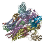
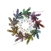
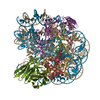
Journal: Nat Commun / Year: 2022 Title: Multiple conformations of trimeric spikes visualized on a non-enveloped virus. Authors: Yinong Zhang / Yanxiang Cui / Jingchen Sun / Z Hong Zhou / Abstract: Many viruses utilize trimeric spikes to gain entry into host cells. However, without in situ structures of these trimeric spikes, a full understanding of this dynamic and essential process of viral ...Many viruses utilize trimeric spikes to gain entry into host cells. However, without in situ structures of these trimeric spikes, a full understanding of this dynamic and essential process of viral infections is not possible. Here we present four in situ and one isolated cryoEM structures of the trimeric spike of the cytoplasmic polyhedrosis virus, a member of the non-enveloped Reoviridae family and a virus historically used as a model in the discoveries of RNA transcription and capping. These structures adopt two drastically different conformations, closed spike and opened spike, which respectively represent the penetration-inactive and penetration-active states. Each spike monomer has four domains: N-terminal, body, claw, and C-terminal. From closed to opened state, the RGD motif-containing C-terminal domain is freed to bind integrins, and the claw domain rotates to expose and project its membrane insertion loops into the cellular membrane. Comparison between turret vertices before and after detachment of the trimeric spike shows that the trimeric spike anchors its N-terminal domain in the iris of the pentameric RNA-capping turret. Sensing of cytosolic S-adenosylmethionine (SAM) and adenosine triphosphate (ATP) by the turret triggers a cascade of events: opening of the iris, detachment of the spike, and initiation of endogenous transcription.
History
Deposition
Dec 31, 2021
-
Header (metadata) release
Feb 2, 2022
-
Map release
Feb 2, 2022
-
Update
Jul 2, 2025
-
Current status
Jul 2, 2025
Processing site: PDBj / Status: Released
-
Structure visualization
Movie
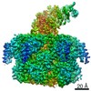
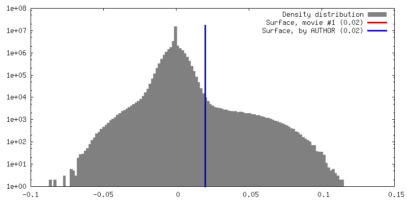
Surface view with section colored by density value
In the structure databanks used in Yorodumi, some data are registered as the other names, "COVID-19 virus" and "2019-nCoV". Here are the details of the virus and the list of structure data.
Jan 31, 2019. EMDB accession codes are about to change! (news from PDBe EMDB page)
EMDB accession codes are about to change! (news from PDBe EMDB page)
The allocation of 4 digits for EMDB accession codes will soon come to an end. Whilst these codes will remain in use, new EMDB accession codes will include an additional digit and will expand incrementally as the available range of codes is exhausted. The current 4-digit format prefixed with “EMD-” (i.e. EMD-XXXX) will advance to a 5-digit format (i.e. EMD-XXXXX), and so on. It is currently estimated that the 4-digit codes will be depleted around Spring 2019, at which point the 5-digit format will come into force.
The EM Navigator/Yorodumi systems omit the EMD- prefix.
Related info.:Q: What is EMD? / ID/Accession-code notation in Yorodumi/EM Navigator
Yorodumi is a browser for structure data from EMDB, PDB, SASBDB, etc.
This page is also the successor to EM Navigator detail page, and also detail information page/front-end page for Omokage search.
The word "yorodu" (or yorozu) is an old Japanese word meaning "ten thousand". "mi" (miru) is to see.
Related info.:EMDB / PDB / SASBDB / Comparison of 3 databanks / Yorodumi Search / Aug 31, 2016. New EM Navigator & Yorodumi / Yorodumi Papers / Jmol/JSmol / Function and homology information / Changes in new EM Navigator and Yorodumi
 Movie
Movie Controller


 Open data
 Basic information
 Map data
 Sample
 Keywords
 Function and homology information
 Bombyx mori cypovirus 1
 Authors
 China,
  United States, 10 items
  Citation
 Structure visualization
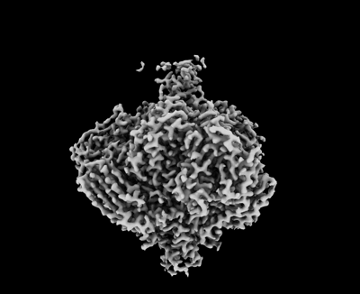
 Downloads & links
 emd_32505.png
 http://ftp.pdbj.org/pub/emdb/structures/EMD-32505


 Z (Sec.)
 Y (Row.)
 X (Col.)


 Sample components
 Processing
 Electron microscopy
 FIELD EMISSION GUN
