Movie
Movie Controller

[English] 日本語
 Yorodumi
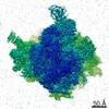
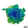
- EMDB-32507: Pentameric turret of Bombyx mori cytoplasmic polyhedrosis virus a... -
+ Open data

- Basic information
| Entry | Database: EMDB / ID: EMD-32507 | |||||||||||||||||||||||||||||||||
|---|---|---|---|---|---|---|---|---|---|---|---|---|---|---|---|---|---|---|---|---|---|---|---|---|---|---|---|---|---|---|---|---|---|---|
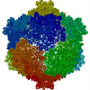
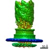
| Title | Pentameric turret of Bombyx mori cytoplasmic polyhedrosis virus after spike detaches. | |||||||||||||||||||||||||||||||||
 Map data Map data | ||||||||||||||||||||||||||||||||||
 Sample Sample |
| |||||||||||||||||||||||||||||||||
 Keywords Keywords | Capping enzyme / VIRAL PROTEIN | |||||||||||||||||||||||||||||||||
| Function / homology | : / : / : / : / Reovirus VP3 protein, guanylyltransferase (GTase) / Reovirus turret protein, bridge domain / Reovirus VP3 protein, Methyltransferase domain 1 / Reovirus VP3 protein, Methyltransferase domain 2 / Structural protein VP3 Function and homology information Function and homology information | |||||||||||||||||||||||||||||||||
| Biological species |   Bombyx mori cypovirus 1 Bombyx mori cypovirus 1 | |||||||||||||||||||||||||||||||||
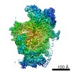
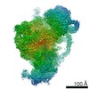
| Method | single particle reconstruction / cryo EM / Resolution: 3.7 Å | |||||||||||||||||||||||||||||||||
 Authors Authors | Zhang Y / Cui Y | |||||||||||||||||||||||||||||||||
| Funding support |  China, China,  United States, 10 items United States, 10 items
| |||||||||||||||||||||||||||||||||
 Citation Citation | Journal: Nat Commun / Year: 2022 Title: Multiple conformations of trimeric spikes visualized on a non-enveloped virus. Authors: Yinong Zhang / Yanxiang Cui / Jingchen Sun / Z Hong Zhou / Abstract: Many viruses utilize trimeric spikes to gain entry into host cells. However, without in situ structures of these trimeric spikes, a full understanding of this dynamic and essential process of viral ...Many viruses utilize trimeric spikes to gain entry into host cells. However, without in situ structures of these trimeric spikes, a full understanding of this dynamic and essential process of viral infections is not possible. Here we present four in situ and one isolated cryoEM structures of the trimeric spike of the cytoplasmic polyhedrosis virus, a member of the non-enveloped Reoviridae family and a virus historically used as a model in the discoveries of RNA transcription and capping. These structures adopt two drastically different conformations, closed spike and opened spike, which respectively represent the penetration-inactive and penetration-active states. Each spike monomer has four domains: N-terminal, body, claw, and C-terminal. From closed to opened state, the RGD motif-containing C-terminal domain is freed to bind integrins, and the claw domain rotates to expose and project its membrane insertion loops into the cellular membrane. Comparison between turret vertices before and after detachment of the trimeric spike shows that the trimeric spike anchors its N-terminal domain in the iris of the pentameric RNA-capping turret. Sensing of cytosolic S-adenosylmethionine (SAM) and adenosine triphosphate (ATP) by the turret triggers a cascade of events: opening of the iris, detachment of the spike, and initiation of endogenous transcription. | |||||||||||||||||||||||||||||||||
| History |
|
- Structure visualization
| Movie |
Movie viewer |
|---|---|
| Structure viewer | EM map: SurfViewMolmilJmol/JSmol |
| Supplemental images |

- Downloads & links
-EMDB archive
| Map data | emd_32507.map.gz | 95.2 MB | EMDB map data format | |
|---|---|---|---|---|
| Header (meta data) | emd-32507-v30.xmlemd-32507.xml | 18.4 KB 18.4 KB | Display Display | EMDB header |
| Images | 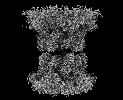 emd_32507.png emd_32507.png | 106 KB | ||
| Filedesc metadata | emd-32507.cif.gz | 6.9 KB | ||
| Archive directory |  http://ftp.pdbj.org/pub/emdb/structures/EMD-32507ftp://ftp.pdbj.org/pub/emdb/structures/EMD-32507 http://ftp.pdbj.org/pub/emdb/structures/EMD-32507ftp://ftp.pdbj.org/pub/emdb/structures/EMD-32507 | HTTPS FTP |
-Related structure data
| Related structure data | 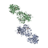 7whpMC  7whmC  7whnC M: atomic model generated by this map C: citing same article ( |
|---|---|
| Similar structure data |


-Links
| EMDB pages | EMDB (EBI/PDBe) / EMDataResource |
|---|
-Map
| File | Download / File: emd_32507.map.gz / Format: CCP4 / Size: 103 MB / Type: IMAGE STORED AS FLOATING POINT NUMBER (4 BYTES) | ||||||||||||||||||||||||||||||||||||||||||||||||||||||||||||||||||||
|---|---|---|---|---|---|---|---|---|---|---|---|---|---|---|---|---|---|---|---|---|---|---|---|---|---|---|---|---|---|---|---|---|---|---|---|---|---|---|---|---|---|---|---|---|---|---|---|---|---|---|---|---|---|---|---|---|---|---|---|---|---|---|---|---|---|---|---|---|---|
| Projections & slices | Image control
Images are generated by Spider. | ||||||||||||||||||||||||||||||||||||||||||||||||||||||||||||||||||||
| Voxel size | X=Y=Z: 1.062 Å | ||||||||||||||||||||||||||||||||||||||||||||||||||||||||||||||||||||
| Density |
| ||||||||||||||||||||||||||||||||||||||||||||||||||||||||||||||||||||
| Symmetry | Space group: 1 | ||||||||||||||||||||||||||||||||||||||||||||||||||||||||||||||||||||
| Details | EMDB XML:
CCP4 map header:
| ||||||||||||||||||||||||||||||||||||||||||||||||||||||||||||||||||||
 Z (Sec.)
 Y (Row.)
 X (Col.)


-Supplemental data
- Sample components
-Entire : Bombyx mori cypovirus 1
| Entire | Name: Bombyx mori cypovirus 1 |
|---|---|
| Components |
|
-Supramolecule #1: Bombyx mori cypovirus 1
| Supramolecule | Name: Bombyx mori cypovirus 1 / type: virus / ID: 1 / Parent: 0 / Macromolecule list: #1 / NCBI-ID: 110829 / Sci species name: Bombyx mori cypovirus 1 / Virus type: VIRION / Virus isolate: STRAIN / Virus enveloped: No / Virus empty: No |
|---|---|
| Host (natural) | Organism:  |
-Macromolecule #1: Structural protein VP3
| Macromolecule | Name: Structural protein VP3 / type: protein_or_peptide / ID: 1 / Number of copies: 2 / Enantiomer: LEVO |
|---|---|
| Source (natural) | Organism: Bombyx mori cypovirus 1 |
| Molecular weight | Theoretical: 120.145797 KDa |
| Sequence | String: MWHYTSINND TRVALDPKPN QIRTITKPNT VPQLGTDYLY TFNSQRRSHT LRLLGPFQYF NFSETDRGHP LFRLPLKYPS KAIPADELI DNLHSWMRSV HLLHVRSEDN TLRYNWMLGV YARSTNYTTP VGQLVVNAPA ILNYSNPQDA FNSVFVALGI D YIDIPITN ...String: MWHYTSINND TRVALDPKPN QIRTITKPNT VPQLGTDYLY TFNSQRRSHT LRLLGPFQYF NFSETDRGHP LFRLPLKYPS KAIPADELI DNLHSWMRSV HLLHVRSEDN TLRYNWMLGV YARSTNYTTP VGQLVVNAPA ILNYSNPQDA FNSVFVALGI D YIDIPITN SNIFDDSSTP YNVRIWHAPT MTEVNHILAL MRKSTLVSTH SSWHWNVLHT FHYRSESDMI DHFAAKILED WR QKEKLDK GALVEADRVI QRLIPLSSST YVQRLAAIGA LYPNEFTENV LDLSRLSTAL LQLSDTYYQH ANDQLRRLYR RMY NDSRTL YMTQRHQELL LAQITADPNI LLYPYTYIFT TIPTSMNYIS NTGQGRIKHS LTVTGATEHD TVADIVLGQT GEDV ITISM VEPMSIAVED MYGYVLDTPT RDIWPADEQI EQKGDAVALY DTKTSRALGM FNNTVRIDDL LSPLLSLVYR TYIKG DTMT MTQGSLDHLT LCAAVDSDIT FVGNRMIAPL PEGYIPKPMH RNNSTMKMLS LYVALKKLEN FATNSYLMAP DTSIIL LGA EREPAVNILR RFNRNVSNVR IIGMGDRAVE PNIRVRVPFP IDKNISADFI ICDINSYEDQ SFESMFSETI SVVTTCA SA ATRALVKINH PSEYMINSVI ERLSQLGGVF YHTALLKTAS QNPYSYETYI YITPIAAAVR FPFYSNSAMI NRYMTAVA D DEMPIIPSIH TVIKGHSNTY SPGLFCGCVD VQSAPLALSQ LKSYCSEATT WRVDSDDNLV NIIARIDPAR IALEFRTRS NTSAYHEYQR YVPNGLGFKV RKTREFRYMH REVTFIHKLM MYALIREQIS LTENMTQVVS IGGRNLADIS VVPLNMKYVV IDPATRIET LTQEKKNIEV QSRPFQFDAA NMDLENNSIY LFIAVIMNEP NGAATPARMQ MDKIRNVATA MLTRTNCVAY I SFYEAGII TRLDQSTAHK TIRVEEGRLK VANYVPVDTL VEADVTLMLR DIGITHEIIR PSTPELIDAC SNYGIRLGST GG AVLDVFN HYSPVIKLVR S UniProtKB: Structural protein VP3 |
-Macromolecule #2: S-ADENOSYLMETHIONINE
| Macromolecule | Name: S-ADENOSYLMETHIONINE / type: ligand / ID: 2 / Number of copies: 4 / Formula: SAM |
|---|---|
| Molecular weight | Theoretical: 398.437 Da |
| Chemical component information |  ChemComp-SAM: |
-Macromolecule #3: ADENOSINE-5'-TRIPHOSPHATE
| Macromolecule | Name: ADENOSINE-5'-TRIPHOSPHATE / type: ligand / ID: 3 / Number of copies: 2 / Formula: ATP |
|---|---|
| Molecular weight | Theoretical: 507.181 Da |
| Chemical component information |  ChemComp-ATP: |
-Experimental details
-Structure determination
| Method | cryo EM |
|---|---|
 Processing Processing | single particle reconstruction |
| Aggregation state | particle |
-Sample preparation
| Buffer | pH: 8 Component:
| ||||||||||||||||||
|---|---|---|---|---|---|---|---|---|---|---|---|---|---|---|---|---|---|---|---|
| Vitrification | Cryogen name: ETHANE-PROPANE |
- Electron microscopy
| Microscope | FEI TITAN KRIOS |
|---|---|
| Image recording | Film or detector model: GATAN K2 QUANTUM (4k x 4k) / Detector mode: SUPER-RESOLUTION / Average electron dose: 40.0 e/Å2 |
| Electron beam | Acceleration voltage: 300 kV / Electron source:  FIELD EMISSION GUN FIELD EMISSION GUN |
| Electron optics | Illumination mode: FLOOD BEAM / Imaging mode: BRIGHT FIELD / Nominal defocus max: 2.3000000000000003 µm / Nominal defocus min: 1.0 µm / Nominal magnification: 130000 |
| Experimental equipment |  Model: Titan Krios / Image courtesy: FEI Company |
+Image processing
-Atomic model buiding 1
| Initial model | PDB ID: Chain - Chain ID: A / Chain - Residue range: 1-1057 / Chain - Source name: PDB / Chain - Initial model type: experimental model |
|---|---|
| Refinement | Space: REAL / Protocol: FLEXIBLE FIT |
| Output model | PDB-7whp: |