National Institutes of Health/National Institute Of Allergy and Infectious Diseases (NIH/NIAID)
75N93019C00074
United States
Citation
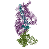
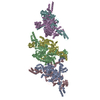
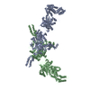
Journal: Cell Host Microbe / Year: 2021 Title: Complete Mapping of Mutations to the SARS-CoV-2 Spike Receptor-Binding Domain that Escape Antibody Recognition. Authors: Allison J Greaney / Tyler N Starr / Pavlo Gilchuk / Seth J Zost / Elad Binshtein / Andrea N Loes / Sarah K Hilton / John Huddleston / Rachel Eguia / Katharine H D Crawford / Adam S Dingens / ...Authors: Allison J Greaney / Tyler N Starr / Pavlo Gilchuk / Seth J Zost / Elad Binshtein / Andrea N Loes / Sarah K Hilton / John Huddleston / Rachel Eguia / Katharine H D Crawford / Adam S Dingens / Rachel S Nargi / Rachel E Sutton / Naveenchandra Suryadevara / Paul W Rothlauf / Zhuoming Liu / Sean P J Whelan / Robert H Carnahan / James E Crowe / Jesse D Bloom / Abstract: Antibodies targeting the SARS-CoV-2 spike receptor-binding domain (RBD) are being developed as therapeutics and are a major contributor to neutralizing antibody responses elicited by infection. Here, ...Antibodies targeting the SARS-CoV-2 spike receptor-binding domain (RBD) are being developed as therapeutics and are a major contributor to neutralizing antibody responses elicited by infection. Here, we describe a deep mutational scanning method to map how all amino-acid mutations in the RBD affect antibody binding and apply this method to 10 human monoclonal antibodies. The escape mutations cluster on several surfaces of the RBD that broadly correspond to structurally defined antibody epitopes. However, even antibodies targeting the same surface often have distinct escape mutations. The complete escape maps predict which mutations are selected during viral growth in the presence of single antibodies. They further enable the design of escape-resistant antibody cocktails-including cocktails of antibodies that compete for binding to the same RBD surface but have different escape mutations. Therefore, complete escape-mutation maps enable rational design of antibody therapeutics and assessment of the antigenic consequences of viral evolution.
History
Deposition
Sep 8, 2020
-
Header (metadata) release
Oct 14, 2020
-
Map release
Oct 14, 2020
-
Update
Jan 27, 2021
-
Current status
Jan 27, 2021
Processing site: RCSB / Status: Released
-
Structure visualization
Movie
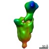
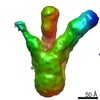
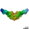
Surface view with section colored by density value
Film or detector model: GATAN ULTRASCAN 4000 (4k x 4k) / Number grids imaged: 1 / Number real images: 237 / Average exposure time: 1.0 sec. / Average electron dose: 30.0 e/Å2
Electron beam
Acceleration voltage: 200 kV / Electron source: FIELD EMISSION GUN
In the structure databanks used in Yorodumi, some data are registered as the other names, "COVID-19 virus" and "2019-nCoV". Here are the details of the virus and the list of structure data.
Jan 31, 2019. EMDB accession codes are about to change! (news from PDBe EMDB page)
EMDB accession codes are about to change! (news from PDBe EMDB page)
The allocation of 4 digits for EMDB accession codes will soon come to an end. Whilst these codes will remain in use, new EMDB accession codes will include an additional digit and will expand incrementally as the available range of codes is exhausted. The current 4-digit format prefixed with “EMD-” (i.e. EMD-XXXX) will advance to a 5-digit format (i.e. EMD-XXXXX), and so on. It is currently estimated that the 4-digit codes will be depleted around Spring 2019, at which point the 5-digit format will come into force.
The EM Navigator/Yorodumi systems omit the EMD- prefix.
Related info.:Q: What is EMD? / ID/Accession-code notation in Yorodumi/EM Navigator
Yorodumi is a browser for structure data from EMDB, PDB, SASBDB, etc.
This page is also the successor to EM Navigator detail page, and also detail information page/front-end page for Omokage search.
The word "yorodu" (or yorozu) is an old Japanese word meaning "ten thousand". "mi" (miru) is to see.
Related info.:EMDB / PDB / SASBDB / Comparison of 3 databanks / Yorodumi Search / Aug 31, 2016. New EM Navigator & Yorodumi / Yorodumi Papers / Jmol/JSmol / Function and homology information / Changes in new EM Navigator and Yorodumi
 Movie
Movie Controller

 Yorodumi
 Open data
 Basic information
 Map data
 Sample
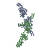
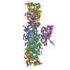
 Severe acute respiratory syndrome coronavirus 2 /
  Homo sapiens (human)
 Authors
 United States, 2 items
  Citation
 Structure visualization
 Movie viewer
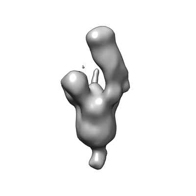
 Downloads & links
 emd_22627.png
 http://ftp.pdbj.org/pub/emdb/structures/EMD-22627
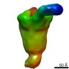

 Z (Sec.)
 Y (Row.)
 X (Col.)


 Sample components
 Processing
 Electron microscopy
 FIELD EMISSION GUN
