 Movie
Movie Controller

[English] 日本語
 Yorodumi
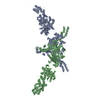
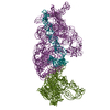
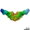
- PDB-5i6i: Crystal structure of a dBCCP-variant of Chaetomium thermophilum a... -
+ Open data

- Basic information
| Entry | Database: PDB / ID: 5i6i | ||||||||||||
|---|---|---|---|---|---|---|---|---|---|---|---|---|---|
| Title | Crystal structure of a dBCCP-variant of Chaetomium thermophilum acetyl-CoA carboxylase | ||||||||||||
 Components Components | Acetyl-CoA carboxylase-like protein,Acetyl-CoA carboxylase-like protein,Acetyl-CoA carboxylase-like protein | ||||||||||||
 Keywords Keywords | LIGASE / Carboxylase / Fatty acid metabolism / Multienzyme / Carrier protein-dependent enzyme | ||||||||||||
| Function / homology |  Function and homology information Function and homology informationmalonyl-CoA biosynthetic process / biotin carboxylase activity / acetyl-CoA carboxylase activity / fatty acid biosynthetic process / mitochondrion / ATP binding / metal ion binding Similarity search - Function | ||||||||||||
| Biological species |  Chaetomium thermophilum (fungus)Chaetomium thermophilum var. thermophilum DSM 1495 (fungus) Chaetomium thermophilum (fungus)Chaetomium thermophilum var. thermophilum DSM 1495 (fungus) | ||||||||||||
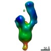
| Method |  X-RAY DIFFRACTION / SYNCHROTRON / MOLECULAR REPLACEMENT / Resolution: 8.4 Å X-RAY DIFFRACTION / SYNCHROTRON / MOLECULAR REPLACEMENT / Resolution: 8.4 Å | ||||||||||||
 Authors Authors | Hunkeler, M. / Stuttfeld, E. / Hagmann, A. / Imseng, S. / Maier, T. | ||||||||||||
| Funding support |  Switzerland, 3items Switzerland, 3items
| ||||||||||||
 Citation Citation | Journal: Nat Commun / Year: 2016 Title: The dynamic organization of fungal acetyl-CoA carboxylase. Authors: Hunkeler, M. / Stuttfeld, E. / Hagmann, A. / Imseng, S. / Maier, T. | ||||||||||||
| History |
|
- Structure visualization
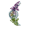
| Structure viewer | Molecule: MolmilJmol/JSmol |
|---|
- Downloads & links
-Download
| PDBx/mmCIF format | 5i6i.cif.gz | 1.2 MB | Display | PDBx/mmCIF format |
|---|---|---|---|---|
| PDB format | pdb5i6i.ent.gz | 982.8 KB | Display | PDB format |
| PDBx/mmJSON format | 5i6i.json.gz | Tree view | PDBx/mmJSON format | |
| Others |  Other downloads Other downloads |
-Validation report
| Arichive directory | https://data.pdbj.org/pub/pdb/validation_reports/i6/5i6iftp://data.pdbj.org/pub/pdb/validation_reports/i6/5i6i | HTTPS FTP |
|---|
-Related structure data
| Related structure data |  5i6eSC  5i6fSC  5i6gC  5i6hC  5i87C S: Starting model for refinement C: citing same article ( |
|---|---|
| Similar structure data |
-Links
 PDBj
PDBj


- Assembly
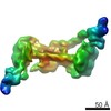
| Deposited unit | 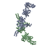
| ||||||||
|---|---|---|---|---|---|---|---|---|---|
| 1 |
| ||||||||
| Unit cell |
|
-Components
| #1: Protein | Mass: 248740.312 Da / Num. of mol.: 2 Source method: isolated from a genetically manipulated source Details: The crystallized construct contains amino acids 1-2297 (aligned to accession code: G0S3L5). The carrier protein (residues 701-766 aligned to accession code G0S3L5) was removed from the ...Details: The crystallized construct contains amino acids 1-2297 (aligned to accession code: G0S3L5). The carrier protein (residues 701-766 aligned to accession code G0S3L5) was removed from the construct and replaced by a GSG linker. Due to formatting restrictions the SEQRES card was truncated C-terminally to residue R2261. C-terminal stretches after L2259 in chain A could not be modeled unambiguously and were interpreted as poly-Ala/UNK residues Source: (gene. exp.) Chaetomium thermophilum (strain DSM 1495 / CBS 144.50 / IMI 039719) (fungus), (gene. exp.) Chaetomium thermophilum var. thermophilum DSM 1495 (fungus)Gene: CTHT_0021690 / Strain: DSM 1495 / CBS 144.50 / IMI 039719 / Production host:   Spodoptera frugiperda (fall armyworm) / References: UniProt: G0S3L5, acetyl-CoA carboxylase Spodoptera frugiperda (fall armyworm) / References: UniProt: G0S3L5, acetyl-CoA carboxylaseHas protein modification | N | |
|---|
-Experimental details
-Experiment
| Experiment | Method: X-RAY DIFFRACTION / Number of used crystals: 1 |
|---|
- Sample preparation
| Crystal | Density Matthews: 6.34 Å3/Da / Density % sol: 80.61 % |
|---|---|
| Crystal grow | Temperature: 292.15 K / Method: vapor diffusion, sitting drop Details: Morpheus buffer system 3, Morpheus ethylene glycol mix, PEG4000, Glycerol PH range: 8.5 |
-Data collection
| Diffraction | Mean temperature: 100 K |
|---|---|
| Diffraction source | Source: SYNCHROTRON / Site: SLS / Beamline: X06SA / Wavelength: 0.99986 Å |
| Detector | Type: DECTRIS PILATUS 6M-F / Detector: PIXEL / Date: Oct 11, 2014 |
| Radiation | Protocol: SINGLE WAVELENGTH / Monochromatic (M) / Laue (L): M / Scattering type: x-ray |
| Radiation wavelength | Wavelength: 0.99986 Å / Relative weight: 1 |
| Reflection | Resolution: 8.4→49.95 Å / Num. obs: 12113 / % possible obs: 99.1 % / Redundancy: 18.5 % / Biso Wilson estimate: 751.38 Å2 / Rmerge(I) obs: 0.294 / Net I/σ(I): 9.05 |
| Reflection shell | Resolution: 8.4→8.62 Å / Redundancy: 18.2 % / Rmerge(I) obs: 3.82 / Mean I/σ(I) obs: 0.9 / % possible all: 99.9 |
- Processing
| Software |
| |||||||||||||||||||||||||||||||||||||||||||||||||||||||||||||||||||||||||||||||||||||||||||||||||||||||||||||||||||||||||||||||||||||||||||||||||||||||||||||||||||||||||||||||||||||||||||||||||||||||||||||||||||||||||||||||||
|---|---|---|---|---|---|---|---|---|---|---|---|---|---|---|---|---|---|---|---|---|---|---|---|---|---|---|---|---|---|---|---|---|---|---|---|---|---|---|---|---|---|---|---|---|---|---|---|---|---|---|---|---|---|---|---|---|---|---|---|---|---|---|---|---|---|---|---|---|---|---|---|---|---|---|---|---|---|---|---|---|---|---|---|---|---|---|---|---|---|---|---|---|---|---|---|---|---|---|---|---|---|---|---|---|---|---|---|---|---|---|---|---|---|---|---|---|---|---|---|---|---|---|---|---|---|---|---|---|---|---|---|---|---|---|---|---|---|---|---|---|---|---|---|---|---|---|---|---|---|---|---|---|---|---|---|---|---|---|---|---|---|---|---|---|---|---|---|---|---|---|---|---|---|---|---|---|---|---|---|---|---|---|---|---|---|---|---|---|---|---|---|---|---|---|---|---|---|---|---|---|---|---|---|---|---|---|---|---|---|---|---|---|---|---|---|---|---|---|---|---|---|---|---|---|---|---|
| Refinement | Method to determine structure: MOLECULAR REPLACEMENT Starting model: 5i6f, 5i6e Resolution: 8.4→49.95 Å / Cor.coef. Fo:Fc: 0.6999 / Cor.coef. Fo:Fc free: 0.6775 / Cross valid method: THROUGHOUT / σ(F): 0 / SU Rfree Blow DPI: 3.654
| |||||||||||||||||||||||||||||||||||||||||||||||||||||||||||||||||||||||||||||||||||||||||||||||||||||||||||||||||||||||||||||||||||||||||||||||||||||||||||||||||||||||||||||||||||||||||||||||||||||||||||||||||||||||||||||||||
| Displacement parameters | Biso mean: 250.2 Å2
| |||||||||||||||||||||||||||||||||||||||||||||||||||||||||||||||||||||||||||||||||||||||||||||||||||||||||||||||||||||||||||||||||||||||||||||||||||||||||||||||||||||||||||||||||||||||||||||||||||||||||||||||||||||||||||||||||
| Refine analyze | Luzzati coordinate error obs: 0 Å | |||||||||||||||||||||||||||||||||||||||||||||||||||||||||||||||||||||||||||||||||||||||||||||||||||||||||||||||||||||||||||||||||||||||||||||||||||||||||||||||||||||||||||||||||||||||||||||||||||||||||||||||||||||||||||||||||
| Refinement step | Cycle: LAST / Resolution: 8.4→49.95 Å
| |||||||||||||||||||||||||||||||||||||||||||||||||||||||||||||||||||||||||||||||||||||||||||||||||||||||||||||||||||||||||||||||||||||||||||||||||||||||||||||||||||||||||||||||||||||||||||||||||||||||||||||||||||||||||||||||||
| Refine LS restraints |
| |||||||||||||||||||||||||||||||||||||||||||||||||||||||||||||||||||||||||||||||||||||||||||||||||||||||||||||||||||||||||||||||||||||||||||||||||||||||||||||||||||||||||||||||||||||||||||||||||||||||||||||||||||||||||||||||||
| LS refinement shell | Resolution: 8.4→9.2 Å / Total num. of bins used: 6
| |||||||||||||||||||||||||||||||||||||||||||||||||||||||||||||||||||||||||||||||||||||||||||||||||||||||||||||||||||||||||||||||||||||||||||||||||||||||||||||||||||||||||||||||||||||||||||||||||||||||||||||||||||||||||||||||||
| Refinement TLS params. | Method: refined / Refine-ID: X-RAY DIFFRACTION
| |||||||||||||||||||||||||||||||||||||||||||||||||||||||||||||||||||||||||||||||||||||||||||||||||||||||||||||||||||||||||||||||||||||||||||||||||||||||||||||||||||||||||||||||||||||||||||||||||||||||||||||||||||||||||||||||||
| Refinement TLS group |
|
