 Movie
Movie Controller

[English] 日本語
 Yorodumi
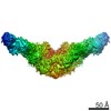
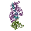
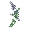
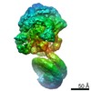
- PDB-6oq5: Structure of the full-length Clostridium difficile toxin B in com... -
+ Open data

- Basic information
| Entry | Database: PDB / ID: 6oq5 | ||||||
|---|---|---|---|---|---|---|---|
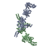
| Title | Structure of the full-length Clostridium difficile toxin B in complex with 3 VHHs | ||||||
 Components Components |
| ||||||
 Keywords Keywords | TOXIN / Toxin VHH | ||||||
| Function / homology |  Function and homology information Function and homology informationhost cell cytosol / glycosyltransferase activity / cysteine-type peptidase activity / host cell endosome membrane / toxin activity / lipid binding / host cell plasma membrane / proteolysis / extracellular region / metal ion binding Similarity search - Function | ||||||
| Biological species |  Clostridioides difficile (bacteria) Clostridioides difficile (bacteria) Camelidae (mammal) Camelidae (mammal) | ||||||
| Method |  X-RAY DIFFRACTION / SYNCHROTRON / MOLECULAR REPLACEMENT / Resolution: 3.87 Å X-RAY DIFFRACTION / SYNCHROTRON / MOLECULAR REPLACEMENT / Resolution: 3.87 Å | ||||||
 Authors Authors | Chen, P. / Lam, K. / Jin, R. | ||||||
| Funding support |  United States, 1items United States, 1items
| ||||||
 Citation Citation | Journal: Nat.Struct.Mol.Biol. / Year: 2019 Title: Structure of the full-length Clostridium difficile toxin B. Authors: Chen, P. / Lam, K.H. / Liu, Z. / Mindlin, F.A. / Chen, B. / Gutierrez, C.B. / Huang, L. / Zhang, Y. / Hamza, T. / Feng, H. / Matsui, T. / Bowen, M.E. / Perry, K. / Jin, R. | ||||||
| History |
|
- Structure visualization
| Structure viewer | Molecule: MolmilJmol/JSmol |
|---|
- Downloads & links
-Download
| PDBx/mmCIF format | 6oq5.cif.gz | 551.5 KB | Display | PDBx/mmCIF format |
|---|---|---|---|---|
| PDB format | pdb6oq5.ent.gz | 437.9 KB | Display | PDB format |
| PDBx/mmJSON format | 6oq5.json.gz | Tree view | PDBx/mmJSON format | |
| Others |  Other downloads Other downloads |
-Validation report
| Arichive directory | https://data.pdbj.org/pub/pdb/validation_reports/oq/6oq5ftp://data.pdbj.org/pub/pdb/validation_reports/oq/6oq5 | HTTPS FTP |
|---|
-Related structure data
| Related structure data |  6oq6C  6oq7C  6oq8C  2bvlS  3peeS 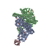 3v0aS  4nc2S  4np4S  4r04S 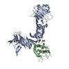 6c0bS S: Starting model for refinement C: citing same article ( |
|---|---|
| Similar structure data |

-Links
 PDBj
PDBj


- Assembly
| Deposited unit | 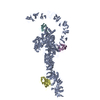
| ||||||||
|---|---|---|---|---|---|---|---|---|---|
| 1 |
| ||||||||
| Unit cell |
|
-Components
-Protein , 1 types, 1 molecules A
| #1: Protein | Mass: 270380.812 Da / Num. of mol.: 1 Source method: isolated from a genetically manipulated source Source: (gene. exp.) Clostridioides difficile (bacteria) / Gene: tcdB / Production host: Bacillus megaterium (bacteria) / References: UniProt: M4NKV9 |
|---|
-Antibody , 3 types, 3 molecules DEF
| #2: Antibody | Mass: 16864.762 Da / Num. of mol.: 1 Source method: isolated from a genetically manipulated source Source: (gene. exp.) Camelidae (mammal) / Production host: |
|---|---|
| #3: Antibody | Mass: 14678.397 Da / Num. of mol.: 1 Source method: isolated from a genetically manipulated source Source: (gene. exp.) Camelidae (mammal) / Production host: |
| #4: Antibody | Mass: 15300.087 Da / Num. of mol.: 1 Source method: isolated from a genetically manipulated source Source: (gene. exp.) Camelidae (mammal) / Production host: |
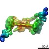
-Non-polymers , 2 types, 2 molecules 

| #5: Chemical | ChemComp-ZN /  Mass: 65.409 Da / Num. of mol.: 1 / Source method: obtained synthetically / Formula: Zn / Feature type: SUBJECT OF INVESTIGATION Mass: 65.409 Da / Num. of mol.: 1 / Source method: obtained synthetically / Formula: Zn / Feature type: SUBJECT OF INVESTIGATION |
|---|---|
| #6: Chemical | ChemComp-MG /  Mass: 24.305 Da / Num. of mol.: 1 / Source method: obtained synthetically / Formula: Mg Mass: 24.305 Da / Num. of mol.: 1 / Source method: obtained synthetically / Formula: Mg |
-Details
| Has protein modification | Y |
|---|
-Experimental details
-Experiment
| Experiment | Method: X-RAY DIFFRACTION / Number of used crystals: 1 |
|---|
- Sample preparation
| Crystal | Density Matthews: 3.58 Å3/Da / Density % sol: 65.61 % |
|---|---|
| Crystal grow | Temperature: 291 K / Method: vapor diffusion, hanging drop Details: 0.1 M sodium acetate, 0.1M magnesium acetate, and 5% PEG 8K (final pH 5.2) |
-Data collection
| Diffraction | Mean temperature: 100 K / Serial crystal experiment: N |
|---|---|
| Diffraction source | Source: SYNCHROTRON / Site: APS / Beamline: 24-ID-C / Wavelength: 0.97918 Å |
| Detector | Type: ADSC QUANTUM 315 / Detector: CCD / Date: Jun 15, 2016 |
| Radiation | Protocol: SINGLE WAVELENGTH / Monochromatic (M) / Laue (L): M / Scattering type: x-ray |
| Radiation wavelength | Wavelength: 0.97918 Å / Relative weight: 1 |
| Reflection | Resolution: 3.87→48.91 Å / Num. obs: 42971 / % possible obs: 99.4 % / Redundancy: 3.7 % / Rmerge(I) obs: 0.137 / Net I/σ(I): 7.7 |
| Reflection shell | Resolution: 3.87→3.97 Å / Rmerge(I) obs: 1.548 / Num. unique obs: 2123 |
- Processing
| Software |
| ||||||||||||||||||||||||||||||||||||||||||||||||||||||||||||||||||||||||||||||||||||||||||||||||||||||||||||||||||||||||||||||||||||||||||||||||||||||||||||||||||||||||||||||||||||||
|---|---|---|---|---|---|---|---|---|---|---|---|---|---|---|---|---|---|---|---|---|---|---|---|---|---|---|---|---|---|---|---|---|---|---|---|---|---|---|---|---|---|---|---|---|---|---|---|---|---|---|---|---|---|---|---|---|---|---|---|---|---|---|---|---|---|---|---|---|---|---|---|---|---|---|---|---|---|---|---|---|---|---|---|---|---|---|---|---|---|---|---|---|---|---|---|---|---|---|---|---|---|---|---|---|---|---|---|---|---|---|---|---|---|---|---|---|---|---|---|---|---|---|---|---|---|---|---|---|---|---|---|---|---|---|---|---|---|---|---|---|---|---|---|---|---|---|---|---|---|---|---|---|---|---|---|---|---|---|---|---|---|---|---|---|---|---|---|---|---|---|---|---|---|---|---|---|---|---|---|---|---|---|---|
| Refinement | Method to determine structure: MOLECULAR REPLACEMENT Starting model: 2BVL, 4R04, 3PEE, 6C0B, 3V0A, 4NP4, and 4NC2 Resolution: 3.87→48.91 Å / Cor.coef. Fo:Fc: 0.933 / Cor.coef. Fo:Fc free: 0.902 / SU B: 81.488 / SU ML: 1.029 / Cross valid method: THROUGHOUT / ESU R Free: 0.903
| ||||||||||||||||||||||||||||||||||||||||||||||||||||||||||||||||||||||||||||||||||||||||||||||||||||||||||||||||||||||||||||||||||||||||||||||||||||||||||||||||||||||||||||||||||||||
| Solvent computation | Ion probe radii: 0.8 Å / Shrinkage radii: 0.8 Å / VDW probe radii: 1.2 Å | ||||||||||||||||||||||||||||||||||||||||||||||||||||||||||||||||||||||||||||||||||||||||||||||||||||||||||||||||||||||||||||||||||||||||||||||||||||||||||||||||||||||||||||||||||||||
| Displacement parameters | Biso mean: 169.399 Å2
| ||||||||||||||||||||||||||||||||||||||||||||||||||||||||||||||||||||||||||||||||||||||||||||||||||||||||||||||||||||||||||||||||||||||||||||||||||||||||||||||||||||||||||||||||||||||
| Refinement step | Cycle: 1 / Resolution: 3.87→48.91 Å
| ||||||||||||||||||||||||||||||||||||||||||||||||||||||||||||||||||||||||||||||||||||||||||||||||||||||||||||||||||||||||||||||||||||||||||||||||||||||||||||||||||||||||||||||||||||||
| Refine LS restraints |
|