 Movie
Movie Controller

[English] 日本語
 Yorodumi
- PDB-7t0r: Crystal structure of the anti-CD4 adnectin 6940_B01 as a complex ... -
+ Open data

- Basic information
| Entry | Database: PDB / ID: 7t0r | ||||||
|---|---|---|---|---|---|---|---|
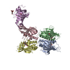
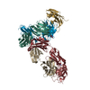
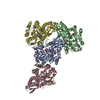
| Title | Crystal structure of the anti-CD4 adnectin 6940_B01 as a complex with the extracellular domains of CD4 and ibalizumab fAb | ||||||
 Components Components |
| ||||||
 Keywords Keywords | ANTIVIRAL PROTEIN / Inhibitor / HIV gp140 / CD4 / adnectin / ibalizumab / HIV Env / combinectin | ||||||
| Function / homology |  Function and homology information Function and homology informationhelper T cell enhancement of adaptive immune response / interleukin-16 binding / interleukin-16 receptor activity / cellular response to ionomycin / maintenance of protein location in cell / response to methamphetamine hydrochloride / T cell selection / MHC class II protein binding / interleukin-15-mediated signaling pathway / cellular response to granulocyte macrophage colony-stimulating factor stimulus ...helper T cell enhancement of adaptive immune response / interleukin-16 binding / interleukin-16 receptor activity / cellular response to ionomycin / maintenance of protein location in cell / response to methamphetamine hydrochloride / T cell selection / MHC class II protein binding / interleukin-15-mediated signaling pathway / cellular response to granulocyte macrophage colony-stimulating factor stimulus / positive regulation of monocyte differentiation / Alpha-defensins / Nef Mediated CD4 Down-regulation / regulation of T cell activation / response to vitamin D / Other interleukin signaling / leukocyte chemotaxis / extracellular matrix structural constituent / T cell receptor complex / enzyme-linked receptor protein signaling pathway / Translocation of ZAP-70 to Immunological synapse / Phosphorylation of CD3 and TCR zeta chains / macrophage differentiation / Generation of second messenger molecules / immunoglobulin binding / T cell differentiation / Co-inhibition by PD-1 / positive regulation of calcium ion transport into cytosol / Binding and entry of HIV virion / coreceptor activity / positive regulation of interleukin-2 production / positive regulation of T cell proliferation / cell surface receptor protein tyrosine kinase signaling pathway / protein tyrosine kinase binding / T cell activation / clathrin-coated endocytic vesicle membrane / Vpu mediated degradation of CD4 / positive regulation of inflammatory response / MHC class II protein complex binding / response to estradiol / transmembrane signaling receptor activity / T cell receptor signaling pathway / Downstream TCR signaling / Cargo recognition for clathrin-mediated endocytosis / Clathrin-mediated endocytosis / virus receptor activity / signaling receptor activity / response to ethanol / adaptive immune response / early endosome / cell surface receptor signaling pathway / cell adhesion / immune response / membrane raft / endoplasmic reticulum lumen / external side of plasma membrane / signaling receptor binding / symbiont entry into host cell / lipid binding / protein kinase binding / endoplasmic reticulum membrane / enzyme binding / signal transduction / protein homodimerization activity / : / zinc ion binding / identical protein binding / plasma membrane Similarity search - Function | ||||||
| Biological species |   Homo sapiens (human) Homo sapiens (human)synthetic construct (others) | ||||||
| Method |  X-RAY DIFFRACTION / SYNCHROTRON / MOLECULAR REPLACEMENT / molecular replacement / Resolution: 3.65 Å X-RAY DIFFRACTION / SYNCHROTRON / MOLECULAR REPLACEMENT / molecular replacement / Resolution: 3.65 Å | ||||||
 Authors Authors | Williams, S.P. / Concha, N.O. / Wensel, D.L. / Hong, X. | ||||||
| Funding support |  United States, 1items United States, 1items
| ||||||
 Citation Citation | Journal: J Mol Biol / Year: 2022 Title: Novel Bent Conformation of CD4 Induced by HIV-1 Inhibitor Indirectly Prevents Productive Viral Attachment. Authors: David Wensel / Shawn Williams / David P Dixon / Paris Ward / Patti McCormick / Nestor Concha / Eugene Stewart / Xuan Hong / Charles Mazzucco / Shreya Pal / Bo Ding / Christoph Fellinger / Mark Krystal /  Abstract: GSK3732394 is a multi-specific biologic inhibitor of HIV entry currently under clinical evaluation. A key component of this molecule is an adnectin (6940_B01) that binds to CD4 and inhibits ...GSK3732394 is a multi-specific biologic inhibitor of HIV entry currently under clinical evaluation. A key component of this molecule is an adnectin (6940_B01) that binds to CD4 and inhibits downstream actions of gp160. Studies were performed to determine the binding site of the adnectin on CD4 and to understand the mechanism of inhibition. Using hydrogen-deuterium exchange with mass spectrometry (HDX), CD4 peptides showed differential rates of deuteration (either enhanced or slowed) in the presence of the adnectin that mapped predominantly to the interface of domains 2 and 3 (D2-D3). In addition, an X-ray crystal structure of an ibalizumab Fab/CD4(D1-D4)/adnectin complex revealed an extensive interface between the adnectin and residues on CD4 domains D2-D4 that stabilize a novel T-shaped CD4 conformation. A cryo-EM map of the gp140/CD4/GSK3732394 complex clearly shows the bent conformation for CD4 while bound to gp140. Mutagenic analyses on CD4 confirmed that amino acid F202 forms a key interaction with the adnectin. In addition, amino acid L151 was shown to be a critical indirect determinant of the specificity for binding to the human CD4 protein over related primate CD4 molecules, as it appears to modulate CD4's flexibility to adopt the adnectin-bound conformation. The significant conformational change of CD4 upon adnectin binding brings the D1 domain of CD4 in proximity to the host cell membrane surface, thereby re-orienting the gp120 binding site in a direction that is inaccessible to incoming virus due to a steric clash between gp160 trimers on the virus surface and the target cell membrane. | ||||||
| History |
|
- Structure visualization
| Structure viewer | Molecule: MolmilJmol/JSmol |
|---|
- Downloads & links
-Download
| PDBx/mmCIF format | 7t0r.cif.gz | 415.2 KB | Display | PDBx/mmCIF format |
|---|---|---|---|---|
| PDB format | pdb7t0r.ent.gz | 268.5 KB | Display | PDB format |
| PDBx/mmJSON format | 7t0r.json.gz | Tree view | PDBx/mmJSON format | |
| Others |  Other downloads Other downloads |
-Validation report
| Arichive directory | https://data.pdbj.org/pub/pdb/validation_reports/t0/7t0rftp://data.pdbj.org/pub/pdb/validation_reports/t0/7t0r | HTTPS FTP |
|---|
-Related structure data
| Related structure data |  7t0oC  3o2dS C: citing same article ( S: Starting model for refinement |
|---|---|
| Similar structure data |


-Links
 PDBj
PDBj


- Assembly
| Deposited unit | 
| ||||||||||||||||||||||||||||||||||||||||||||||||||||||||||||||||||||||||||||||||||||||||||||||||||||||||||||||||||||||||||||||||||
|---|---|---|---|---|---|---|---|---|---|---|---|---|---|---|---|---|---|---|---|---|---|---|---|---|---|---|---|---|---|---|---|---|---|---|---|---|---|---|---|---|---|---|---|---|---|---|---|---|---|---|---|---|---|---|---|---|---|---|---|---|---|---|---|---|---|---|---|---|---|---|---|---|---|---|---|---|---|---|---|---|---|---|---|---|---|---|---|---|---|---|---|---|---|---|---|---|---|---|---|---|---|---|---|---|---|---|---|---|---|---|---|---|---|---|---|---|---|---|---|---|---|---|---|---|---|---|---|---|---|---|---|
| 1 | 
| ||||||||||||||||||||||||||||||||||||||||||||||||||||||||||||||||||||||||||||||||||||||||||||||||||||||||||||||||||||||||||||||||||
| 2 | 
| ||||||||||||||||||||||||||||||||||||||||||||||||||||||||||||||||||||||||||||||||||||||||||||||||||||||||||||||||||||||||||||||||||
| Unit cell |
| ||||||||||||||||||||||||||||||||||||||||||||||||||||||||||||||||||||||||||||||||||||||||||||||||||||||||||||||||||||||||||||||||||
| Components on special symmetry positions |
| ||||||||||||||||||||||||||||||||||||||||||||||||||||||||||||||||||||||||||||||||||||||||||||||||||||||||||||||||||||||||||||||||||
| Noncrystallographic symmetry (NCS) | NCS domain:
NCS domain segments:
NCS ensembles :
|
-Components
-Protein , 2 types, 4 molecules ADGI
| #3: Protein | Mass: 41667.727 Da / Num. of mol.: 2 / Fragment: Extra-cellular domains D1-D4 Source method: isolated from a genetically manipulated source Source: (gene. exp.) Homo sapiens (human) / Gene: CD4 / Cell line (production host): HEK / Production host: Homo sapiens (human) / References: UniProt: P01730#4: Protein | Mass: 10314.485 Da / Num. of mol.: 2 Source method: isolated from a genetically manipulated source Source: (gene. exp.) synthetic construct (others) / Production host:  |
|---|
-Antibody , 2 types, 4 molecules LBHC
| #1: Antibody | Mass: 24245.900 Da / Num. of mol.: 2 Source method: isolated from a genetically manipulated source Source: (gene. exp.)  Cricetulus griseus (Chinese hamster) Cricetulus griseus (Chinese hamster)#2: Antibody | Mass: 24785.529 Da / Num. of mol.: 2 Source method: isolated from a genetically manipulated source Source: (gene. exp.) Cricetulus griseus (Chinese hamster) |
|---|
-Sugars / Non-polymers , 2 types, 3 molecules 

| #5: Sugar |  Type: D-saccharide, beta linking / Mass: 221.208 Da / Num. of mol.: 2 / Source method: obtained synthetically / Formula: C8H15NO6 Type: D-saccharide, beta linking / Mass: 221.208 Da / Num. of mol.: 2 / Source method: obtained synthetically / Formula: C8H15NO6#6: Chemical | ChemComp-SO4 / |  Mass: 96.063 Da / Num. of mol.: 1 / Source method: obtained synthetically / Formula: SO4 Mass: 96.063 Da / Num. of mol.: 1 / Source method: obtained synthetically / Formula: SO4 |
|---|
-Details
| Has ligand of interest | N |
|---|---|
| Has protein modification | Y |
-Experimental details
-Experiment
| Experiment | Method: X-RAY DIFFRACTION / Number of used crystals: 1 |
|---|
- Sample preparation
| Crystal | Density Matthews: 2.83 Å3/Da / Density % sol: 56.56 % |
|---|---|
| Crystal grow | Temperature: 295 K / Method: vapor diffusion, hanging drop Details: 0.01 M Na3 Citrate, 15.5% PEG3350, 0.3 M (NH4)2H Citrate, 0.1 M AmSO4 PH range: 7.0 - 7.6 / Temp details: room temperature |
-Data collection
| Diffraction | Mean temperature: 270 K / Ambient temp details: Throughout / Serial crystal experiment: N | |||||||||||||||||||||||||||||||||||||||||||||||||||||||||||||||||||||||||||||||||||||||||||||||||||||||||||||||||||||||||||||||||||||||||||||||||||||||||||||||||||||||||||||||||||||||||||||
|---|---|---|---|---|---|---|---|---|---|---|---|---|---|---|---|---|---|---|---|---|---|---|---|---|---|---|---|---|---|---|---|---|---|---|---|---|---|---|---|---|---|---|---|---|---|---|---|---|---|---|---|---|---|---|---|---|---|---|---|---|---|---|---|---|---|---|---|---|---|---|---|---|---|---|---|---|---|---|---|---|---|---|---|---|---|---|---|---|---|---|---|---|---|---|---|---|---|---|---|---|---|---|---|---|---|---|---|---|---|---|---|---|---|---|---|---|---|---|---|---|---|---|---|---|---|---|---|---|---|---|---|---|---|---|---|---|---|---|---|---|---|---|---|---|---|---|---|---|---|---|---|---|---|---|---|---|---|---|---|---|---|---|---|---|---|---|---|---|---|---|---|---|---|---|---|---|---|---|---|---|---|---|---|---|---|---|---|---|---|---|
| Diffraction source | Source: SYNCHROTRON / Site: APS / Beamline: 22-ID / Wavelength: 0.987 Å | |||||||||||||||||||||||||||||||||||||||||||||||||||||||||||||||||||||||||||||||||||||||||||||||||||||||||||||||||||||||||||||||||||||||||||||||||||||||||||||||||||||||||||||||||||||||||||||
| Detector | Type: DECTRIS EIGER X 16M / Detector: PIXEL / Date: Mar 29, 2019 | |||||||||||||||||||||||||||||||||||||||||||||||||||||||||||||||||||||||||||||||||||||||||||||||||||||||||||||||||||||||||||||||||||||||||||||||||||||||||||||||||||||||||||||||||||||||||||||
| Radiation | Monochromator: Si(111) / Protocol: SINGLE WAVELENGTH / Monochromatic (M) / Laue (L): M / Scattering type: x-ray | |||||||||||||||||||||||||||||||||||||||||||||||||||||||||||||||||||||||||||||||||||||||||||||||||||||||||||||||||||||||||||||||||||||||||||||||||||||||||||||||||||||||||||||||||||||||||||||
| Radiation wavelength | Wavelength: 0.987 Å / Relative weight: 1 | |||||||||||||||||||||||||||||||||||||||||||||||||||||||||||||||||||||||||||||||||||||||||||||||||||||||||||||||||||||||||||||||||||||||||||||||||||||||||||||||||||||||||||||||||||||||||||||
| Reflection | Resolution: 3.5→100 Å / Num. obs: 27334 / % possible obs: 92.2 % / Redundancy: 4.7 % / Biso Wilson estimate: 70.88 Å2 / CC1/2: 0.98 / CC star: 0.995 / Rmerge(I) obs: 0.196 / Rpim(I) all: 0.098 / Rrim(I) all: 0.22 / Χ2: 0.91 / Net I/σ(I): 4.6 | |||||||||||||||||||||||||||||||||||||||||||||||||||||||||||||||||||||||||||||||||||||||||||||||||||||||||||||||||||||||||||||||||||||||||||||||||||||||||||||||||||||||||||||||||||||||||||||
| Reflection shell | Diffraction-ID: 1
|
-Phasing
| Phasing | Method: molecular replacement | ||||||
|---|---|---|---|---|---|---|---|
| Phasing MR | R rigid body: 0.476
|
- Processing
| Software |
| ||||||||||||||||||||||||||||||||||||||||||||||||||||||||||||||||||||||||||||||||||||
|---|---|---|---|---|---|---|---|---|---|---|---|---|---|---|---|---|---|---|---|---|---|---|---|---|---|---|---|---|---|---|---|---|---|---|---|---|---|---|---|---|---|---|---|---|---|---|---|---|---|---|---|---|---|---|---|---|---|---|---|---|---|---|---|---|---|---|---|---|---|---|---|---|---|---|---|---|---|---|---|---|---|---|---|---|---|
| Refinement | Method to determine structure: MOLECULAR REPLACEMENT Starting model: 3O2D Resolution: 3.65→29.53 Å / SU ML: 0.4947 / Cross valid method: FREE R-VALUE / σ(F): 1.35 / Phase error: 28.4107 Stereochemistry target values: GeoStd + Monomer Library + CDL v1.2
| ||||||||||||||||||||||||||||||||||||||||||||||||||||||||||||||||||||||||||||||||||||
| Solvent computation | Shrinkage radii: 0.9 Å / VDW probe radii: 1.11 Å / Solvent model: FLAT BULK SOLVENT MODEL | ||||||||||||||||||||||||||||||||||||||||||||||||||||||||||||||||||||||||||||||||||||
| Displacement parameters | Biso mean: 69 Å2 | ||||||||||||||||||||||||||||||||||||||||||||||||||||||||||||||||||||||||||||||||||||
| Refinement step | Cycle: LAST / Resolution: 3.65→29.53 Å
| ||||||||||||||||||||||||||||||||||||||||||||||||||||||||||||||||||||||||||||||||||||
| Refine LS restraints |
| ||||||||||||||||||||||||||||||||||||||||||||||||||||||||||||||||||||||||||||||||||||
| Refine LS restraints NCS |
| ||||||||||||||||||||||||||||||||||||||||||||||||||||||||||||||||||||||||||||||||||||
| LS refinement shell |
|
