Mass: 18.015 Da / Num. of mol.: 215 / Source method: isolated from a natural source / Formula: H2O
Has protein modification
Y
-
Experimental details
-
Experiment
Experiment
Method: X-RAY DIFFRACTION / Number of used crystals: 1
-
Sample preparation
Crystal
Density Matthews: 3.31 Å3/Da / Density % sol: 62.86 %
Crystal grow
Temperature: 293 K / Method: vapor diffusion, hanging drop / pH: 5.5 Details: 3 M Na malonate, pH 5.5, VAPOR DIFFUSION, HANGING DROP, temperature 293.0K
Monochromator: Cryo-Cooled Si(111) double crystal / Protocol: SINGLE WAVELENGTH / Monochromatic (M) / Laue (L): M / Scattering type: x-ray
Radiation wavelength
Wavelength: 0.9793 Å / Relative weight: 1
Reflection
Resolution: 2.19→48.6 Å / Num. all: 48479 / Num. obs: 45445 / % possible obs: 93.5 %
Reflection shell
Resolution: 2.19→2.25 Å / Redundancy: 6.4 % / Mean I/σ(I) obs: 2.7 / Num. unique all: 2836 / Rsym value: 0.631 / % possible all: 89.3
-
Processing
Software
Name
Version
Classification
ADSC
Quantum
datacollection
PHASER
phasing
REFMAC
5.5.0109
refinement
HKL-2000
datareduction
HKL-2000
datascaling
Refinement
Method to determine structure: MOLECULAR REPLACEMENT / Resolution: 2.19→48.6 Å / Cor.coef. Fo:Fc: 0.952 / Cor.coef. Fo:Fc free: 0.931 / SU B: 11.181 / SU ML: 0.126 / Cross valid method: THROUGHOUT / ESU R Free: 0.174 / Stereochemistry target values: MAXIMUM LIKELIHOOD / Details: HYDROGENS HAVE BEEN ADDED IN THE RIDING POSITIONS
Rfactor
Num. reflection
% reflection
Selection details
Rfree
0.2242
2295
5.1 %
RANDOM
Rwork
0.19206
-
-
-
obs
0.19369
43149
93.74 %
-
all
-
45445
-
-
Solvent computation
Ion probe radii: 0.8 Å / Shrinkage radii: 0.8 Å / VDW probe radii: 1.4 Å / Solvent model: MASK
Displacement parameters
Biso mean: 44.913 Å2
Baniso -1
Baniso -2
Baniso -3
1-
-0.6 Å2
0 Å2
-0 Å2
2-
-
0.96 Å2
0 Å2
3-
-
-
-0.37 Å2
Refinement step
Cycle: LAST / Resolution: 2.19→48.6 Å
Protein
Nucleic acid
Ligand
Solvent
Total
Num. atoms
4751
0
0
215
4966
Refine LS restraints
Refine-ID
Type
Dev ideal
Dev ideal target
Number
X-RAY DIFFRACTION
r_bond_refined_d
0.015
0.022
4858
X-RAY DIFFRACTION
r_bond_other_d
X-RAY DIFFRACTION
r_angle_refined_deg
1.572
1.957
6599
X-RAY DIFFRACTION
r_angle_other_deg
X-RAY DIFFRACTION
r_dihedral_angle_1_deg
6.621
5
613
X-RAY DIFFRACTION
r_dihedral_angle_2_deg
38.311
25.101
198
X-RAY DIFFRACTION
r_dihedral_angle_3_deg
17.174
15
831
X-RAY DIFFRACTION
r_dihedral_angle_4_deg
20.326
15
17
X-RAY DIFFRACTION
r_chiral_restr
0.105
0.2
747
X-RAY DIFFRACTION
r_gen_planes_refined
0.007
0.021
3623
X-RAY DIFFRACTION
r_gen_planes_other
X-RAY DIFFRACTION
r_nbd_refined
X-RAY DIFFRACTION
r_nbd_other
X-RAY DIFFRACTION
r_nbtor_refined
X-RAY DIFFRACTION
r_nbtor_other
X-RAY DIFFRACTION
r_xyhbond_nbd_refined
X-RAY DIFFRACTION
r_xyhbond_nbd_other
X-RAY DIFFRACTION
r_metal_ion_refined
X-RAY DIFFRACTION
r_metal_ion_other
X-RAY DIFFRACTION
r_symmetry_vdw_refined
X-RAY DIFFRACTION
r_symmetry_vdw_other
X-RAY DIFFRACTION
r_symmetry_hbond_refined
X-RAY DIFFRACTION
r_symmetry_hbond_other
X-RAY DIFFRACTION
r_symmetry_metal_ion_refined
X-RAY DIFFRACTION
r_symmetry_metal_ion_other
X-RAY DIFFRACTION
r_mcbond_it
0.897
1.5
3065
X-RAY DIFFRACTION
r_mcbond_other
X-RAY DIFFRACTION
r_mcangle_it
1.73
2
4970
X-RAY DIFFRACTION
r_scbond_it
2.582
3
1793
X-RAY DIFFRACTION
r_scangle_it
4.268
4.5
1629
X-RAY DIFFRACTION
r_rigid_bond_restr
X-RAY DIFFRACTION
r_sphericity_free
X-RAY DIFFRACTION
r_sphericity_bonded
LS refinement shell
Resolution: 2.19→2.252 Å / Total num. of bins used: 20
Rfactor
Num. reflection
% reflection
Rfree
0.28
152
-
Rwork
0.259
2836
-
obs
-
-
85.35 %
Refinement TLS params.
Method: refined / Refine-ID: X-RAY DIFFRACTION
ID
L11 (°2)
L12 (°2)
L13 (°2)
L22 (°2)
L23 (°2)
L33 (°2)
S11 (Å °)
S12 (Å °)
S13 (Å °)
S21 (Å °)
S22 (Å °)
S23 (Å °)
S31 (Å °)
S32 (Å °)
S33 (Å °)
T11 (Å2)
T12 (Å2)
T13 (Å2)
T22 (Å2)
T23 (Å2)
T33 (Å2)
Origin x (Å)
Origin y (Å)
Origin z (Å)
1
1.7315
-0.0186
1.3527
0.2476
0.0358
1.7138
0.0243
-0.0949
0.0709
0.1214
-0.0726
0.0081
0.0215
-0.1703
0.0483
0.1293
-0.0236
-0.0006
0.2684
0.0082
0.0872
3.758
15.592
57.388
2
2.116
-0.5813
1.3705
0.3255
-0.6698
1.5394
0.2106
0.4124
-0.1605
0.0191
-0.1495
0.0115
0.1109
0.298
-0.061
0.1553
0.0304
-0.0305
0.3242
-0.0174
0.0974
13.251
5.862
46.16
3
2.642
1.2648
-0.7443
1.1762
0.1488
2.7514
-0.0456
0.6024
-0.0487
-0.1448
0.0767
0.1012
0.1514
-0.0872
-0.0311
0.0759
0.0514
-0.047
0.731
-0.0352
0.043
-12.896
9.347
12.078
Refinement TLS group
ID
Refine-ID
Refine TLS-ID
Auth asym-ID
Auth seq-ID
1
X-RAY DIFFRACTION
1
L
-10 - 9999
2
X-RAY DIFFRACTION
2
H
-10 - 9999
3
X-RAY DIFFRACTION
3
A
-10 - 9999
+
About Yorodumi
-
News
-
Feb 9, 2022. New format data for meta-information of EMDB entries
New format data for meta-information of EMDB entries
Version 3 of the EMDB header file is now the official format.
The previous official version 1.9 will be removed from the archive.
In the structure databanks used in Yorodumi, some data are registered as the other names, "COVID-19 virus" and "2019-nCoV". Here are the details of the virus and the list of structure data.
Jan 31, 2019. EMDB accession codes are about to change! (news from PDBe EMDB page)
EMDB accession codes are about to change! (news from PDBe EMDB page)
The allocation of 4 digits for EMDB accession codes will soon come to an end. Whilst these codes will remain in use, new EMDB accession codes will include an additional digit and will expand incrementally as the available range of codes is exhausted. The current 4-digit format prefixed with “EMD-” (i.e. EMD-XXXX) will advance to a 5-digit format (i.e. EMD-XXXXX), and so on. It is currently estimated that the 4-digit codes will be depleted around Spring 2019, at which point the 5-digit format will come into force.
The EM Navigator/Yorodumi systems omit the EMD- prefix.
Related info.:Q: What is EMD? / ID/Accession-code notation in Yorodumi/EM Navigator
Yorodumi is a browser for structure data from EMDB, PDB, SASBDB, etc.
This page is also the successor to EM Navigator detail page, and also detail information page/front-end page for Omokage search.
The word "yorodu" (or yorozu) is an old Japanese word meaning "ten thousand". "mi" (miru) is to see.
Related info.:EMDB / PDB / SASBDB / Comparison of 3 databanks / Yorodumi Search / Aug 31, 2016. New EM Navigator & Yorodumi / Yorodumi Papers / Jmol/JSmol / Function and homology information / Changes in new EM Navigator and Yorodumi
 Movie
Movie Controller

 Yorodumi
 Open data
 Basic information
 Components
 Keywords
 Function and homology information
 Homo sapiens (human)
 X-RAY DIFFRACTION /
  Authors
 Citation
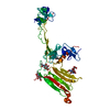
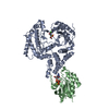
 Structure visualization
 Downloads & links
 Other downloads


 PDBj
PDBj


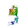
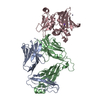
 Assembly

 Spodoptera frugiperda (fall armyworm) / References: UniProt: P01730
 Mass: 18.015 Da / Num. of mol.: 215 / Source method: isolated from a natural source / Formula: H2O
 Sample preparation
 / Beamline: 24-ID-C / Wavelength: 0.9793 Å
 Processing