 Movie
Movie Controller

[English] 日本語
 Yorodumi
- PDB-4udu: Crystal structure of staphylococcal enterotoxin E in complex with... -
+ Open data

- Basic information
| Entry | Database: PDB / ID: 4udu | ||||||
|---|---|---|---|---|---|---|---|
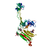
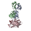
| Title | Crystal structure of staphylococcal enterotoxin E in complex with a T cell receptor | ||||||
 Components Components |
| ||||||
 Keywords Keywords | IMMUNE SYSTEM / SUPERANTIGEN / T CELL RECEPTOR / MAJOR HISTOCOMPATIBILITY COMPLEX | ||||||
| Function / homology |  Function and homology information Function and homology informationalpha-beta T cell receptor complex / T cell receptor complex / Translocation of ZAP-70 to Immunological synapse / Phosphorylation of CD3 and TCR zeta chains / alpha-beta T cell activation / Generation of second messenger molecules / Co-inhibition by PD-1 / response to bacterium / Immunoregulatory interactions between a Lymphoid and a non-Lymphoid cell / T cell receptor signaling pathway ...alpha-beta T cell receptor complex / T cell receptor complex / Translocation of ZAP-70 to Immunological synapse / Phosphorylation of CD3 and TCR zeta chains / alpha-beta T cell activation / Generation of second messenger molecules / Co-inhibition by PD-1 / response to bacterium / Immunoregulatory interactions between a Lymphoid and a non-Lymphoid cell / T cell receptor signaling pathway / Downstream TCR signaling / toxin activity / adaptive immune response / extracellular region / metal ion binding / plasma membrane Similarity search - Function | ||||||
| Biological species |  HOMO SAPIENS (human) HOMO SAPIENS (human)  STAPHYLOCOCCUS AUREUS (bacteria) STAPHYLOCOCCUS AUREUS (bacteria) | ||||||
| Method |  X-RAY DIFFRACTION / SYNCHROTRON / MOLECULAR REPLACEMENT / Resolution: 2.5 Å X-RAY DIFFRACTION / SYNCHROTRON / MOLECULAR REPLACEMENT / Resolution: 2.5 Å | ||||||
 Authors Authors | Rodstrom, K.E.J. / Regenthal, P. / Lindkvist-Petersson, K. | ||||||
 Citation Citation | Journal: Plos One / Year: 2015 Title: Structure of Staphylococcal Enterotoxin E in Complex with Tcr Defines the Role of Tcr Loop Positioning in Superantigen Recognition. Authors: Rodstrom, K.E.J. / Regenthal, P. / Lindkvist-Petersson, K. | ||||||
| History |
|
- Structure visualization
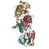
| Structure viewer | Molecule: MolmilJmol/JSmol |
|---|
- Downloads & links
-Download
| PDBx/mmCIF format | 4udu.cif.gz | 262.6 KB | Display | PDBx/mmCIF format |
|---|---|---|---|---|
| PDB format | pdb4udu.ent.gz | 211.1 KB | Display | PDB format |
| PDBx/mmJSON format | 4udu.json.gz | Tree view | PDBx/mmJSON format | |
| Others |  Other downloads Other downloads |
-Validation report
| Arichive directory | https://data.pdbj.org/pub/pdb/validation_reports/ud/4uduftp://data.pdbj.org/pub/pdb/validation_reports/ud/4udu | HTTPS FTP |
|---|
-Related structure data
| Related structure data |  4udtSC  1esfS S: Starting model for refinement C: citing same article ( |
|---|---|
| Similar structure data |


-Links
 PDBj
PDBj


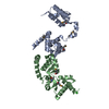
- Assembly
| Deposited unit | 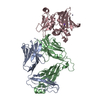
| ||||||||
|---|---|---|---|---|---|---|---|---|---|
| 1 |
| ||||||||
| Unit cell |
|
-Components
-Protein , 3 types, 3 molecules ABC
| #1: Protein | Mass: 22812.125 Da / Num. of mol.: 1 Fragment: VARIABLE DOMAIN TRAV22, RESIDUES 1-112, CONSTANT DOMAIN TRAC1, RESIDUES 2-95 Mutation: YES Source method: isolated from a genetically manipulated source Source: (gene. exp.) HOMO SAPIENS (human) / Cell: T-LYMPHOCYTE / Gene: TRAC, TCRA / Production host: |
|---|---|
| #2: Protein | Mass: 27765.832 Da / Num. of mol.: 1 Fragment: VARIABLE DOMAIN TRBV7-9, RESIDUES 20-115, CONSTANT DOMAIN TRBC2, RESIDUES 1-129 Mutation: YES Source method: isolated from a genetically manipulated source Source: (gene. exp.) HOMO SAPIENS (human) / Cell: T-LYMPHOCYTE / Gene: TCRBV6S4A1, TRBV7-9, TRBC2, TCRBC2 / Production host: |
| #3: Protein | Mass: 26806.947 Da / Num. of mol.: 1 / Fragment: OB DOMAIN AND BETA GRASP DOMAIN, RESIDUES 25-257 Source method: isolated from a genetically manipulated source Source: (gene. exp.) STAPHYLOCOCCUS AUREUS (bacteria) / Gene: ENTE / Production host: |
-Non-polymers , 3 types, 53 molecules 


| #4: Chemical | ChemComp-ZN /  Mass: 65.409 Da / Num. of mol.: 1 / Source method: obtained synthetically / Formula: Zn Mass: 65.409 Da / Num. of mol.: 1 / Source method: obtained synthetically / Formula: Zn | ||
|---|---|---|---|
| #5: Chemical |  Mass: 22.990 Da / Num. of mol.: 2 / Source method: obtained synthetically / Formula: Na Mass: 22.990 Da / Num. of mol.: 2 / Source method: obtained synthetically / Formula: Na#6: Water | ChemComp-HOH / | Mass: 18.015 Da / Num. of mol.: 50 / Source method: isolated from a natural source / Formula: H2O |
-Details
| Has protein modification | Y |
|---|---|
| Sequence details | THE STRUCTURE CONTAINS EXTRACELLULAR TCR ALPHA VARIABLE AND CONSTANT DOMAINS. DATABASE REFERENCE IS ...THE STRUCTURE CONTAINS EXTRACELLU |
-Experimental details
-Experiment
| Experiment | Method: X-RAY DIFFRACTION / Number of used crystals: 1 |
|---|
- Sample preparation
| Crystal | Density Matthews: 2.9 Å3/Da / Density % sol: 58 % |
|---|---|
| Crystal grow | pH: 9 Details: 15% W/V PEG 20000, 0.1 M GLYCINE PH 9.0, 0.1 M NACL |
-Data collection
| Diffraction | Mean temperature: 100 K |
|---|---|
| Diffraction source | Source: SYNCHROTRON / Site: ESRF  / Beamline: ID23-1 / Wavelength: 0.97628 / Beamline: ID23-1 / Wavelength: 0.97628 |
| Detector | Type: ADSC QUANTUM 315 / Detector: CCD / Date: Oct 6, 2012 / Details: TOROIDAL MIRRORS |
| Radiation | Monochromator: SI(111) / Protocol: SINGLE WAVELENGTH / Monochromatic (M) / Laue (L): M / Scattering type: x-ray |
| Radiation wavelength | Wavelength: 0.97628 Å / Relative weight: 1 |
| Reflection | Resolution: 2.5→47.81 Å / Num. obs: 31731 / % possible obs: 99.5 % / Observed criterion σ(I): 2 / Redundancy: 4.4 % / Biso Wilson estimate: 66.64 Å2 / Rmerge(I) obs: 0.08 / Net I/σ(I): 10.6 |
| Reflection shell | Resolution: 2.5→2.6 Å / Redundancy: 4.4 % / Rmerge(I) obs: 0.77 / Mean I/σ(I) obs: 2 / % possible all: 98.2 |
- Processing
| Software |
| ||||||||||||||||||||||||||||||||||||||||||||||||||||||||||||||||||||||||||||||||||||||||||||||||||||||||||||||||||
|---|---|---|---|---|---|---|---|---|---|---|---|---|---|---|---|---|---|---|---|---|---|---|---|---|---|---|---|---|---|---|---|---|---|---|---|---|---|---|---|---|---|---|---|---|---|---|---|---|---|---|---|---|---|---|---|---|---|---|---|---|---|---|---|---|---|---|---|---|---|---|---|---|---|---|---|---|---|---|---|---|---|---|---|---|---|---|---|---|---|---|---|---|---|---|---|---|---|---|---|---|---|---|---|---|---|---|---|---|---|---|---|---|---|---|---|
| Refinement | Method to determine structure: MOLECULAR REPLACEMENT Starting model: SUPERANTIGEN FROM PDB ENTRY 1ESF AND TCR FROM PDB ENTRY 4UDT Resolution: 2.5→32.33 Å / Cor.coef. Fo:Fc: 0.901 / Cor.coef. Fo:Fc free: 0.8942 / SU R Cruickshank DPI: 0.406 / Cross valid method: THROUGHOUT / σ(F): 0 / SU R Blow DPI: 0.399 / SU Rfree Blow DPI: 0.251 / SU Rfree Cruickshank DPI: 0.255 Details: REFMAC 5.7, CNS 1.3, BUSTER 2. DISORDERED REGIONS WERE OMITTED FROM THE MODEL.
| ||||||||||||||||||||||||||||||||||||||||||||||||||||||||||||||||||||||||||||||||||||||||||||||||||||||||||||||||||
| Displacement parameters | Biso mean: 66.02 Å2
| ||||||||||||||||||||||||||||||||||||||||||||||||||||||||||||||||||||||||||||||||||||||||||||||||||||||||||||||||||
| Refine analyze | Luzzati coordinate error obs: 0.521 Å | ||||||||||||||||||||||||||||||||||||||||||||||||||||||||||||||||||||||||||||||||||||||||||||||||||||||||||||||||||
| Refinement step | Cycle: LAST / Resolution: 2.5→32.33 Å
| ||||||||||||||||||||||||||||||||||||||||||||||||||||||||||||||||||||||||||||||||||||||||||||||||||||||||||||||||||
| Refine LS restraints |
| ||||||||||||||||||||||||||||||||||||||||||||||||||||||||||||||||||||||||||||||||||||||||||||||||||||||||||||||||||
| LS refinement shell | Resolution: 2.5→2.58 Å / Total num. of bins used: 16
| ||||||||||||||||||||||||||||||||||||||||||||||||||||||||||||||||||||||||||||||||||||||||||||||||||||||||||||||||||
| Refinement TLS params. | Method: refined / Refine-ID: X-RAY DIFFRACTION
| ||||||||||||||||||||||||||||||||||||||||||||||||||||||||||||||||||||||||||||||||||||||||||||||||||||||||||||||||||
| Refinement TLS group |
|
