 Movie
Movie Controller


+ Open data

- Basic information
| Entry | Database: PDB / ID: 6vlw | ||||||
|---|---|---|---|---|---|---|---|
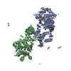
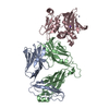
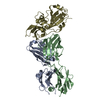
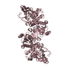
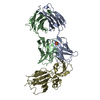
| Title | Crystal Structure of 426cOD in Complex with VRC01 Fab | ||||||
 Components Components |
| ||||||
 Keywords Keywords | IMMUNE SYSTEM/VIRAL PROTEIN / HIV / 426c / Outer Domain / VRC01 / 426cOD / FAB / CD4 / antibody / immunogen / IMMUNE SYSTEM / IMMUNE SYSTEM-VIRAL PROTEIN complex | ||||||
| Function / homology | Immunoglobulins / Immunoglobulin-like / Sandwich / Mainly Beta Function and homology information Function and homology information | ||||||
| Biological species |  Homo sapiens (human) Homo sapiens (human)  Human immunodeficiency virus 1 Human immunodeficiency virus 1 | ||||||
| Method |  X-RAY DIFFRACTION / SYNCHROTRON / MOLECULAR REPLACEMENT / Resolution: 3.42 Å X-RAY DIFFRACTION / SYNCHROTRON / MOLECULAR REPLACEMENT / Resolution: 3.42 Å | ||||||
 Authors Authors | Weidle, C. / Pancera, M. | ||||||
| Funding support |  United States, 1items United States, 1items
| ||||||
 Citation Citation | Journal: Immunity / Year: 2020 Title: HIV-1 VRC01 Germline-Targeting Immunogens Select Distinct Epitope-Specific B Cell Receptors. Authors: Lin, Y.R. / Parks, K.R. / Weidle, C. / Naidu, A.S. / Khechaduri, A. / Riker, A.O. / Takushi, B. / Chun, J.H. / Borst, A.J. / Veesler, D. / Stuart, A. / Agrawal, P. / Gray, M. / Pancera, M. / ...Authors: Lin, Y.R. / Parks, K.R. / Weidle, C. / Naidu, A.S. / Khechaduri, A. / Riker, A.O. / Takushi, B. / Chun, J.H. / Borst, A.J. / Veesler, D. / Stuart, A. / Agrawal, P. / Gray, M. / Pancera, M. / Huang, P.S. / Stamatatos, L. | ||||||
| History |
|
- Structure visualization
| Structure viewer | Molecule: MolmilJmol/JSmol |
|---|
- Downloads & links
-Download
| PDBx/mmCIF format | 6vlw.cif.gz | 239 KB | Display | PDBx/mmCIF format |
|---|---|---|---|---|
| PDB format | pdb6vlw.ent.gz | 176.8 KB | Display | PDB format |
| PDBx/mmJSON format | 6vlw.json.gz | Tree view | PDBx/mmJSON format | |
| Others |  Other downloads Other downloads |
-Validation report
| Arichive directory | https://data.pdbj.org/pub/pdb/validation_reports/vl/6vlwftp://data.pdbj.org/pub/pdb/validation_reports/vl/6vlw | HTTPS FTP |
|---|
-Related structure data
| Related structure data |  5ifaS S: Starting model for refinement |
|---|---|
| Similar structure data |


-Links
 PDBj
PDBj


- Assembly
| Deposited unit | 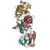
| ||||||||||||
|---|---|---|---|---|---|---|---|---|---|---|---|---|---|
| 1 |
| ||||||||||||
| Unit cell |
|
-Components
-Protein , 1 types, 1 molecules G
| #3: Protein | Mass: 21872.441 Da / Num. of mol.: 1 Source method: isolated from a genetically manipulated source Source: (gene. exp.) Human immunodeficiency virus 1 / Production host: Homo sapiens (human) |
|---|
-Antibody , 2 types, 2 molecules HL
| #1: Antibody | Mass: 24726.000 Da / Num. of mol.: 1 Source method: isolated from a genetically manipulated source Source: (gene. exp.) Homo sapiens (human) / Cell line (production host): HEK293 / Production host: Homo sapiens (human) |
|---|---|
| #2: Antibody | Mass: 23102.570 Da / Num. of mol.: 1 Source method: isolated from a genetically manipulated source Source: (gene. exp.) Homo sapiens (human) / Cell line (production host): HEK293 / Production host: Homo sapiens (human) |
-Sugars , 2 types, 4 molecules 
| #4: Polysaccharide | beta-D-mannopyranose-(1-4)-2-acetamido-2-deoxy-beta-D-glucopyranose-(1-4)-2-acetamido-2-deoxy-beta- ...beta-D-mannopyranose-(1-4)-2-acetamido-2-deoxy-beta-D-glucopyranose-(1-4)-2-acetamido-2-deoxy-beta-D-glucopyranose Source method: isolated from a genetically manipulated source |
|---|---|
| #9: Sugar |  Type: D-saccharide, beta linking / Mass: 221.208 Da / Num. of mol.: 3 Type: D-saccharide, beta linking / Mass: 221.208 Da / Num. of mol.: 3Source method: isolated from a genetically manipulated source Formula: C8H15NO6 |
-Non-polymers , 5 types, 72 molecules 


| #5: Chemical | ChemComp-EDO /  Mass: 62.068 Da / Num. of mol.: 1 / Source method: obtained synthetically / Formula: C2H6O2 Mass: 62.068 Da / Num. of mol.: 1 / Source method: obtained synthetically / Formula: C2H6O2 | ||||||
|---|---|---|---|---|---|---|---|
| #6: Chemical | ChemComp-CL /  Mass: 35.453 Da / Num. of mol.: 8 Mass: 35.453 Da / Num. of mol.: 8Source method: isolated from a genetically manipulated source Formula: Cl #7: Chemical |  Mass: 96.063 Da / Num. of mol.: 2 / Source method: obtained synthetically / Formula: SO4 Mass: 96.063 Da / Num. of mol.: 2 / Source method: obtained synthetically / Formula: SO4#8: Chemical | ChemComp-NA / |  Mass: 22.990 Da / Num. of mol.: 1 Mass: 22.990 Da / Num. of mol.: 1Source method: isolated from a genetically manipulated source Formula: Na #10: Water | ChemComp-HOH / | Mass: 18.015 Da / Num. of mol.: 60 / Source method: isolated from a natural source / Formula: H2O |
-Details
| Has ligand of interest | N |
|---|---|
| Has protein modification | Y |
-Experimental details
-Experiment
| Experiment | Method: X-RAY DIFFRACTION / Number of used crystals: 1 |
|---|
- Sample preparation
| Crystal | Density Matthews: 4.68 Å3/Da / Density % sol: 73.72 % |
|---|---|
| Crystal grow | Temperature: 293 K / Method: vapor diffusion, hanging drop / pH: 6.5 Details: 18.425% PEG 3350, 11.011% 2-Methyl-2,4-pentanediol, 0.22M Lithium Sulfate, 0.11M Imidazole pH 6.5 |
-Data collection
| Diffraction | Mean temperature: 100 K / Serial crystal experiment: N |
|---|---|
| Diffraction source | Source: SYNCHROTRON / Site: ALS / Beamline: 5.0.1 / Wavelength: 1 Å |
| Detector | Type: DECTRIS PILATUS 6M / Detector: PIXEL / Date: Apr 25, 2019 |
| Radiation | Protocol: SINGLE WAVELENGTH / Monochromatic (M) / Laue (L): M / Scattering type: x-ray |
| Radiation wavelength | Wavelength: 1 Å / Relative weight: 1 |
| Reflection | Resolution: 3.42→50 Å / Num. obs: 15995 / % possible obs: 100 % / Redundancy: 6.7 % / Biso Wilson estimate: 63.14 Å2 / CC1/2: 0.992 / Net I/σ(I): 25.57 |
| Reflection shell | Resolution: 3.42→3.48 Å / Num. unique obs: 854 / CC1/2: 0.661 |
- Processing
| Software |
| |||||||||||||||||||||||||||||||||||||||||||||||||
|---|---|---|---|---|---|---|---|---|---|---|---|---|---|---|---|---|---|---|---|---|---|---|---|---|---|---|---|---|---|---|---|---|---|---|---|---|---|---|---|---|---|---|---|---|---|---|---|---|---|---|
| Refinement | Method to determine structure: MOLECULAR REPLACEMENT Starting model: 5IFA Resolution: 3.42→48.57 Å / SU ML: 0.5125 / Cross valid method: FREE R-VALUE / σ(F): 1.34 / Phase error: 27.675 Stereochemistry target values: GeoStd + Monomer Library + CDL v1.2
| |||||||||||||||||||||||||||||||||||||||||||||||||
| Solvent computation | Shrinkage radii: 0.9 Å / VDW probe radii: 1.11 Å / Solvent model: FLAT BULK SOLVENT MODEL | |||||||||||||||||||||||||||||||||||||||||||||||||
| Displacement parameters | Biso mean: 59.24 Å2 | |||||||||||||||||||||||||||||||||||||||||||||||||
| Refinement step | Cycle: LAST / Resolution: 3.42→48.57 Å
| |||||||||||||||||||||||||||||||||||||||||||||||||
| Refine LS restraints |
| |||||||||||||||||||||||||||||||||||||||||||||||||
| LS refinement shell |
|
