 Movie
Movie Controller

[English] 日本語
 Yorodumi
- PDB-1qyy: Crystal Structure of N-Terminal Domain of Human Platelet Receptor... -
+ Open data

- Basic information
| Entry | Database: PDB / ID: 1qyy | |||||||||
|---|---|---|---|---|---|---|---|---|---|---|
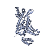
| Title | Crystal Structure of N-Terminal Domain of Human Platelet Receptor Glycoprotein Ib-alpha at 2.8 Angstrom Resolution | |||||||||
 Components Components | Platelet glycoprotein Ib alpha chain | |||||||||
 Keywords Keywords | CELL ADHESION / Platelet Receptors / Glycocalicin / Leucine Rich Repeats | |||||||||
| Function / homology |  Function and homology information Function and homology informationthrombin-activated receptor activity / Enhanced binding of GP1BA variant to VWF multimer:collagen / Defective binding of VWF variant to GPIb:IX:V / blood coagulation, intrinsic pathway / Defective F9 activation / Platelet Adhesion to exposed collagen / positive regulation of platelet activation / megakaryocyte development / GP1b-IX-V activation signalling / regulation of blood coagulation ...thrombin-activated receptor activity / Enhanced binding of GP1BA variant to VWF multimer:collagen / Defective binding of VWF variant to GPIb:IX:V / blood coagulation, intrinsic pathway / Defective F9 activation / Platelet Adhesion to exposed collagen / positive regulation of platelet activation / megakaryocyte development / GP1b-IX-V activation signalling / regulation of blood coagulation / Platelet Aggregation (Plug Formation) / fibrinolysis / : / release of sequestered calcium ion into cytosol / RUNX1 regulates genes involved in megakaryocyte differentiation and platelet function / platelet activation / blood coagulation / signaling receptor activity / cell surface receptor signaling pathway / cell adhesion / external side of plasma membrane / cell surface / extracellular exosome / membrane / plasma membrane Similarity search - Function | |||||||||
| Biological species |  Homo sapiens (human) Homo sapiens (human) | |||||||||
| Method |  X-RAY DIFFRACTION / SYNCHROTRON / MAD / Resolution: 2.8 Å X-RAY DIFFRACTION / SYNCHROTRON / MAD / Resolution: 2.8 Å | |||||||||
 Authors Authors | Varughese, K.I. / Ruggeri, Z.M. / Celikel, R. | |||||||||
 Citation Citation | Journal: Acta Crystallogr.,Sect.D / Year: 2004 Title: Platinum-induced space-group transformation in crystals of the platelet glycoprotein Ib alpha N-terminal domain. Authors: Varughese, K.I. / Ruggeri, Z.M. / Celikel, R. | |||||||||
| History |
|
- Structure visualization
| Structure viewer | Molecule: MolmilJmol/JSmol |
|---|
- Downloads & links
-Download
| PDBx/mmCIF format | 1qyy.cif.gz | 110 KB | Display | PDBx/mmCIF format |
|---|---|---|---|---|
| PDB format | pdb1qyy.ent.gz | 85.5 KB | Display | PDB format |
| PDBx/mmJSON format | 1qyy.json.gz | Tree view | PDBx/mmJSON format | |
| Others |  Other downloads Other downloads |
-Validation report
| Arichive directory | https://data.pdbj.org/pub/pdb/validation_reports/qy/1qyyftp://data.pdbj.org/pub/pdb/validation_reports/qy/1qyy | HTTPS FTP |
|---|
-Related structure data
| Related structure data | |
|---|---|
| Similar structure data |


-Links
 PDBj
PDBj


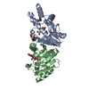
- Assembly
| Deposited unit | 
| ||||||||
|---|---|---|---|---|---|---|---|---|---|
| 1 | 
| ||||||||
| 2 | 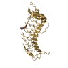
| ||||||||
| Unit cell |
| ||||||||
| Details | Chain A or Chain G represent the Biological unit. |
-Components
| #1: Protein | Mass: 32306.787 Da / Num. of mol.: 2 / Fragment: N-Terminal Domain / Mutation: C65A Source method: isolated from a genetically manipulated source Source: (gene. exp.) Homo sapiens (human) / Gene: GP1BA / Production host:  #2: Polysaccharide | alpha-D-mannopyranose-(1-3)-alpha-D-mannopyranose-(1-4)-2-acetamido-2-deoxy-beta-D-glucopyranose-(1- ...alpha-D-mannopyranose-(1-3)-alpha-D-mannopyranose-(1-4)-2-acetamido-2-deoxy-beta-D-glucopyranose-(1-4)-2-acetamido-2-deoxy-beta-D-glucopyranose | Source method: isolated from a genetically manipulated source #3: Chemical |   Mass: 195.078 Da / Num. of mol.: 3 / Source method: obtained synthetically / Formula: Pt Mass: 195.078 Da / Num. of mol.: 3 / Source method: obtained synthetically / Formula: Pt#4: Sugar | ChemComp-NAG / |   Type: D-saccharide, beta linking / Mass: 221.208 Da / Num. of mol.: 1 Type: D-saccharide, beta linking / Mass: 221.208 Da / Num. of mol.: 1Source method: isolated from a genetically manipulated source Formula: C8H15NO6 #5: Water | ChemComp-HOH / |  Mass: 18.015 Da / Num. of mol.: 56 / Source method: isolated from a natural source / Formula: H2O Mass: 18.015 Da / Num. of mol.: 56 / Source method: isolated from a natural source / Formula: H2OHas protein modification | Y | |
|---|
-Experimental details
-Experiment
| Experiment | Method: X-RAY DIFFRACTION / Number of used crystals: 1 |
|---|
- Sample preparation
| Crystal | Density Matthews: 2.54 Å3/Da / Density % sol: 51.63 % | ||||||||||||||||||||||||
|---|---|---|---|---|---|---|---|---|---|---|---|---|---|---|---|---|---|---|---|---|---|---|---|---|---|
| Crystal grow | Temperature: 277 K / Method: vapor diffusion, hanging drop / pH: 5.6 Details: PEG 6000, SODIUM NITRATE, SODIUM ACETATE, pH 5.6, VAPOR DIFFUSION, HANGING DROP, temperature 277K | ||||||||||||||||||||||||
| Crystal grow | *PLUS Method: vapor diffusion, hanging drop | ||||||||||||||||||||||||
| Components of the solutions | *PLUS
|
-Data collection
| Diffraction | Mean temperature: 100 K | |||||||||
|---|---|---|---|---|---|---|---|---|---|---|
| Diffraction source | Source: SYNCHROTRON / Site: SSRL  / Beamline: BL1-5 / Wavelength: 1.071564, 1.0079675 / Beamline: BL1-5 / Wavelength: 1.071564, 1.0079675 | |||||||||
| Detector | Type: ADSC QUANTUM 4 / Detector: CCD / Date: May 31, 2002 | |||||||||
| Radiation | Monochromator: 2-Crystal Monochromator / Protocol: MAD / Monochromatic (M) / Laue (L): M / Scattering type: x-ray | |||||||||
| Radiation wavelength |
| |||||||||
| Reflection | Resolution: 2.7→50 Å / Num. all: 16932 / Num. obs: 16932 / % possible obs: 94.6 % / Observed criterion σ(F): 0 / Observed criterion σ(I): 0 / Redundancy: 7 % / Rsym value: 0.069 / Net I/σ(I): 8.7 | |||||||||
| Reflection shell | Resolution: 2.7→2.85 Å / Mean I/σ(I) obs: 2.2 / Num. unique all: 1163 / Rsym value: 0.317 / % possible all: 72.2 | |||||||||
| Reflection | *PLUS Highest resolution: 2.7 Å / Num. measured all: 118489 / Rmerge(I) obs: 0.07 | |||||||||
| Reflection shell | *PLUS Highest resolution: 2.7 Å / % possible obs: 72.2 % / Num. unique obs: 1889 / Rmerge(I) obs: 0.317 / Mean I/σ(I) obs: 2.2 |
- Processing
| Software |
| |||||||||||||||||||||||||
|---|---|---|---|---|---|---|---|---|---|---|---|---|---|---|---|---|---|---|---|---|---|---|---|---|---|---|
| Refinement | Method to determine structure: MAD Starting model: Ab initio Resolution: 2.8→15 Å / Cross valid method: R free / σ(F): 0 / σ(I): 0 / Stereochemistry target values: Engh & Huber
| |||||||||||||||||||||||||
| Refinement step | Cycle: LAST / Resolution: 2.8→15 Å
| |||||||||||||||||||||||||
| Refine LS restraints |
| |||||||||||||||||||||||||
| Refinement | *PLUS Rfactor Rfree: 0.2821 / Rfactor Rwork: 0.2201 | |||||||||||||||||||||||||
| Solvent computation | *PLUS | |||||||||||||||||||||||||
| Displacement parameters | *PLUS | |||||||||||||||||||||||||
| LS refinement shell | *PLUS Rfactor Rfree: 0.377 / Rfactor Rwork: 0.318 |
