 Movie
Movie Controller

[English] 日本語
 Yorodumi
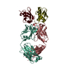
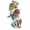
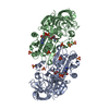
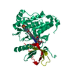
- PDB-4ma7: Crystal structure of mouse prion protein complexed with Promazine -
+ Open data

- Basic information
| Entry | Database: PDB / ID: 4ma7 | ||||||
|---|---|---|---|---|---|---|---|
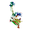
| Title | Crystal structure of mouse prion protein complexed with Promazine | ||||||
 Components Components |
| ||||||
 Keywords Keywords | IMMUNE SYSTEM / Immunoglobulin fold / Fab / Antibody / Mouse prion protein | ||||||
| Function / homology |  Function and homology information Function and homology informationInsertion of tail-anchored proteins into the endoplasmic reticulum membrane / negative regulation of amyloid precursor protein catabolic process / regulation of glutamate receptor signaling pathway / lamin binding / aspartic-type endopeptidase inhibitor activity / regulation of calcium ion import across plasma membrane / glycosaminoglycan binding / regulation of potassium ion transmembrane transport / positive regulation of glutamate receptor signaling pathway / cupric ion binding ...Insertion of tail-anchored proteins into the endoplasmic reticulum membrane / negative regulation of amyloid precursor protein catabolic process / regulation of glutamate receptor signaling pathway / lamin binding / aspartic-type endopeptidase inhibitor activity / regulation of calcium ion import across plasma membrane / glycosaminoglycan binding / regulation of potassium ion transmembrane transport / positive regulation of glutamate receptor signaling pathway / cupric ion binding / negative regulation of interleukin-17 production / ATP-dependent protein binding / type 5 metabotropic glutamate receptor binding / nucleobase-containing compound metabolic process / negative regulation of dendritic spine maintenance / negative regulation of calcineurin-NFAT signaling cascade / negative regulation of interleukin-2 production / response to copper ion / negative regulation of activated T cell proliferation / negative regulation of amyloid-beta formation / response to amyloid-beta / negative regulation of type II interferon production / cuprous ion binding / negative regulation of long-term synaptic potentiation / intracellular copper ion homeostasis / positive regulation of protein targeting to membrane / negative regulation of T cell receptor signaling pathway / response to cadmium ion / side of membrane / positive regulation of calcium-mediated signaling / neuron projection maintenance / inclusion body / cellular response to copper ion / molecular function activator activity / positive regulation of protein localization to plasma membrane / molecular condensate scaffold activity / tubulin binding / cellular response to xenobiotic stimulus / protein homooligomerization / protein destabilization / cellular response to amyloid-beta / terminal bouton / positive regulation of neuron apoptotic process / regulation of protein localization / amyloid-beta binding / protein-folding chaperone binding / signaling receptor activity / protease binding / response to oxidative stress / nuclear membrane / microtubule binding / molecular adaptor activity / transmembrane transporter binding / learning or memory / mitochondrial outer membrane / postsynaptic density / intracellular signal transduction / membrane raft / copper ion binding / dendrite / negative regulation of apoptotic process / protein-containing complex binding / Golgi apparatus / negative regulation of transcription by RNA polymerase II / cell surface / endoplasmic reticulum / membrane / metal ion binding / identical protein binding / plasma membrane / cytosol Similarity search - Function | ||||||
| Biological species |  | ||||||
| Method |  X-RAY DIFFRACTION / SYNCHROTRON / MOLECULAR REPLACEMENT / Resolution: 1.97 Å X-RAY DIFFRACTION / SYNCHROTRON / MOLECULAR REPLACEMENT / Resolution: 1.97 Å | ||||||
 Authors Authors | Baral, P.K. / Swayampakula, M. / James, M.N.G. | ||||||
 Citation Citation | Journal: Structure / Year: 2014 Title: Structural basis of prion inhibition by phenothiazine compounds. Authors: Baral, P.K. / Swayampakula, M. / Rout, M.K. / Kav, N.N. / Spyracopoulos, L. / Aguzzi, A. / James, M.N. | ||||||
| History |
|
- Structure visualization
| Structure viewer | Molecule: MolmilJmol/JSmol |
|---|
- Downloads & links
-Download
| PDBx/mmCIF format | 4ma7.cif.gz | 128.2 KB | Display | PDBx/mmCIF format |
|---|---|---|---|---|
| PDB format | pdb4ma7.ent.gz | 97.5 KB | Display | PDB format |
| PDBx/mmJSON format | 4ma7.json.gz | Tree view | PDBx/mmJSON format | |
| Others |  Other downloads Other downloads |
-Validation report
| Arichive directory | https://data.pdbj.org/pub/pdb/validation_reports/ma/4ma7ftp://data.pdbj.org/pub/pdb/validation_reports/ma/4ma7 | HTTPS FTP |
|---|
-Related structure data
| Related structure data | 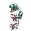 4ma8C  4h88S C: citing same article ( S: Starting model for refinement |
|---|---|
| Similar structure data |


-Links
 PDBj
PDBj


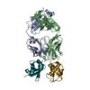
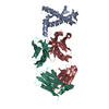
- Assembly
| Deposited unit | 
| ||||||||
|---|---|---|---|---|---|---|---|---|---|
| 1 |
| ||||||||
| Unit cell |
|
-Components
| #1: Protein | Mass: 13360.877 Da / Num. of mol.: 1 / Fragment: UNP residues 116-229 Source method: isolated from a genetically manipulated source Source: (gene. exp.)  |
|---|---|
| #2: Antibody | Mass: 23397.078 Da / Num. of mol.: 1 / Fragment: Fab Source method: isolated from a genetically manipulated source Details: hybridoma / Source: (gene. exp.) |
| #3: Antibody | Mass: 23509.701 Da / Num. of mol.: 1 / Fragment: Fab Source method: isolated from a genetically manipulated source Details: hybridoma / Source: (gene. exp.) |
| #4: Chemical | ChemComp-P2Z /   Mass: 284.419 Da / Num. of mol.: 1 / Source method: obtained synthetically / Formula: C17H20N2S Mass: 284.419 Da / Num. of mol.: 1 / Source method: obtained synthetically / Formula: C17H20N2S |
| #5: Water | ChemComp-HOH /  Mass: 18.015 Da / Num. of mol.: 409 / Source method: isolated from a natural source / Formula: H2O Mass: 18.015 Da / Num. of mol.: 409 / Source method: isolated from a natural source / Formula: H2O |
| Has protein modification | Y |
-Experimental details
-Experiment
| Experiment | Method: X-RAY DIFFRACTION / Number of used crystals: 1 |
|---|
- Sample preparation
| Crystal | Density Matthews: 2.76 Å3/Da / Density % sol: 55.41 % |
|---|---|
| Crystal grow | Temperature: 298 K / Method: vapor diffusion, sitting drop / pH: 6.5 Details: 25% PEG3350, 0.1 M Bis-Tris, pH 6.5, 0.2 M lithium sulfate, VAPOR DIFFUSION, SITTING DROP, temperature 298K |
-Data collection
| Diffraction | Mean temperature: 100 K |
|---|---|
| Diffraction source | Source: SYNCHROTRON / Site: SSRL  / Beamline: BL9-2 / Wavelength: 0.97946 Å / Beamline: BL9-2 / Wavelength: 0.97946 Å |
| Detector | Type: MARMOSAIC 325 mm CCD / Detector: CCD / Date: Feb 4, 2011 |
| Radiation | Monochromator: Double crystal Si(111) / Protocol: SINGLE WAVELENGTH / Monochromatic (M) / Laue (L): M / Scattering type: x-ray |
| Radiation wavelength | Wavelength: 0.97946 Å / Relative weight: 1 |
| Reflection | Resolution: 1.9→50 Å / Num. obs: 52155 / % possible obs: 98.3 % / Observed criterion σ(F): 0 / Observed criterion σ(I): -3 / Redundancy: 4.1 % / Rmerge(I) obs: 0.09 / Net I/σ(I): 15.4 |
| Reflection shell | Resolution: 1.9→1.97 Å / Redundancy: 3.2 % / Rmerge(I) obs: 0.62 / Mean I/σ(I) obs: 2 / % possible all: 85.1 |
- Processing
| Software |
| ||||||||||||||||||||||||||||||||||||||||||||||||||||||||||||||||||||||||||||||||||||||||||||||||||||||||||||||||||||||||||||||||||||||||||||||||||||||||||||||||||||||||||||||||||||||
|---|---|---|---|---|---|---|---|---|---|---|---|---|---|---|---|---|---|---|---|---|---|---|---|---|---|---|---|---|---|---|---|---|---|---|---|---|---|---|---|---|---|---|---|---|---|---|---|---|---|---|---|---|---|---|---|---|---|---|---|---|---|---|---|---|---|---|---|---|---|---|---|---|---|---|---|---|---|---|---|---|---|---|---|---|---|---|---|---|---|---|---|---|---|---|---|---|---|---|---|---|---|---|---|---|---|---|---|---|---|---|---|---|---|---|---|---|---|---|---|---|---|---|---|---|---|---|---|---|---|---|---|---|---|---|---|---|---|---|---|---|---|---|---|---|---|---|---|---|---|---|---|---|---|---|---|---|---|---|---|---|---|---|---|---|---|---|---|---|---|---|---|---|---|---|---|---|---|---|---|---|---|---|---|
| Refinement | Method to determine structure: MOLECULAR REPLACEMENT Starting model: PDB ENTRY 4H88 Resolution: 1.97→34.88 Å / Cor.coef. Fo:Fc: 0.946 / Cor.coef. Fo:Fc free: 0.935 / SU B: 4.154 / SU ML: 0.117 / Cross valid method: THROUGHOUT / ESU R: 0.188 / ESU R Free: 0.163 / Stereochemistry target values: MAXIMUM LIKELIHOOD / Details: HYDROGENS HAVE BEEN ADDED IN THE RIDING POSITIONS
| ||||||||||||||||||||||||||||||||||||||||||||||||||||||||||||||||||||||||||||||||||||||||||||||||||||||||||||||||||||||||||||||||||||||||||||||||||||||||||||||||||||||||||||||||||||||
| Solvent computation | Ion probe radii: 0.8 Å / Shrinkage radii: 0.8 Å / VDW probe radii: 1.2 Å / Solvent model: MASK | ||||||||||||||||||||||||||||||||||||||||||||||||||||||||||||||||||||||||||||||||||||||||||||||||||||||||||||||||||||||||||||||||||||||||||||||||||||||||||||||||||||||||||||||||||||||
| Displacement parameters | Biso mean: 40.636 Å2
| ||||||||||||||||||||||||||||||||||||||||||||||||||||||||||||||||||||||||||||||||||||||||||||||||||||||||||||||||||||||||||||||||||||||||||||||||||||||||||||||||||||||||||||||||||||||
| Refinement step | Cycle: LAST / Resolution: 1.97→34.88 Å
| ||||||||||||||||||||||||||||||||||||||||||||||||||||||||||||||||||||||||||||||||||||||||||||||||||||||||||||||||||||||||||||||||||||||||||||||||||||||||||||||||||||||||||||||||||||||
| Refine LS restraints |
|