 Movie
Movie Controller


+ Open data

- Basic information
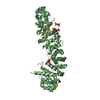
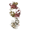
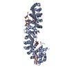
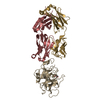
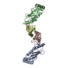
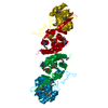
| Entry | Database: PDB / ID: 1dl5 | ||||||
|---|---|---|---|---|---|---|---|
| Title | PROTEIN-L-ISOASPARTATE O-METHYLTRANSFERASE | ||||||
 Components Components | PROTEIN-L-ISOASPARTATE O-METHYLTRANSFERASE | ||||||
 Keywords Keywords | TRANSFERASE / METHYLTRANSFERASE / ISOASPARTYL RESIDUES / PROTEIN REPAIR / DEAMIDATION / POST-TRANSLATIONAL MODIFICATION | ||||||
| Function / homology |  Function and homology information Function and homology informationprotein-L-isoaspartate(D-aspartate) O-methyltransferase / protein-L-isoaspartate (D-aspartate) O-methyltransferase activity / protein repair / methylation / cytoplasm Similarity search - Function | ||||||
| Biological species |   Thermotoga maritima (bacteria) Thermotoga maritima (bacteria) | ||||||
| Method |  X-RAY DIFFRACTION / MIR / Resolution: 1.8 Å X-RAY DIFFRACTION / MIR / Resolution: 1.8 Å | ||||||
 Authors Authors | Skinner, M.M. / Puvathingal, J.M. / Walter, R.L. / Friedman, A.M. | ||||||
 Citation Citation | Journal: Structure Fold.Des. / Year: 2000 Title: Crystal structure of protein isoaspartyl methyltransferase: a catalyst for protein repair. Authors: Skinner, M.M. / Puvathingal, J.M. / Walter, R.L. / Friedman, A.M. | ||||||
| History |
|
- Structure visualization
| Structure viewer | Molecule: MolmilJmol/JSmol |
|---|
- Downloads & links
-Download
| PDBx/mmCIF format | 1dl5.cif.gz | 153.6 KB | Display | PDBx/mmCIF format |
|---|---|---|---|---|
| PDB format | pdb1dl5.ent.gz | 118.5 KB | Display | PDB format |
| PDBx/mmJSON format | 1dl5.json.gz | Tree view | PDBx/mmJSON format | |
| Others |  Other downloads Other downloads |
-Validation report
| Arichive directory | https://data.pdbj.org/pub/pdb/validation_reports/dl/1dl5ftp://data.pdbj.org/pub/pdb/validation_reports/dl/1dl5 | HTTPS FTP |
|---|
-Related structure data
| Similar structure data |
|---|

-Links
 PDBj
PDBj- Assembly
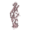
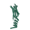
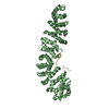
| Deposited unit | 
| ||||||||
|---|---|---|---|---|---|---|---|---|---|
| 1 |
| ||||||||
| 2 | 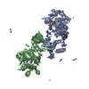
| ||||||||
| Unit cell |
| ||||||||
| Details | THE CRYSTALLOGRAPHIC ASYMMETRIC UNIT IS A DIMER (CHAIN A AND B) RELATED BY THE NON-CRYSTALLOGRAPHIC SYMMETRY OPERATOR. FUNCTIONAL OLIGOMERIZATION STATE IS UNKNOWN. |
-Components
| #1: Protein | Mass: 36440.836 Da / Num. of mol.: 2 Source method: isolated from a genetically manipulated source Source: (gene. exp.) Thermotoga maritima (bacteria) / Plasmid: PET30B / Production host: References: UniProt: Q56308, protein-L-isoaspartate(D-aspartate) O-methyltransferase #2: Chemical | ChemComp-CD /   Mass: 112.411 Da / Num. of mol.: 9 / Source method: obtained synthetically / Formula: Cd / Details: COMMERCIAL SOURCE (SIGMA ) Mass: 112.411 Da / Num. of mol.: 9 / Source method: obtained synthetically / Formula: Cd / Details: COMMERCIAL SOURCE (SIGMA )#3: Chemical | ChemComp-CL /   Mass: 35.453 Da / Num. of mol.: 10 / Source method: obtained synthetically / Formula: Cl Mass: 35.453 Da / Num. of mol.: 10 / Source method: obtained synthetically / Formula: Cl#4: Chemical |   Mass: 384.411 Da / Num. of mol.: 2 / Source method: obtained synthetically / Formula: C14H20N6O5S Mass: 384.411 Da / Num. of mol.: 2 / Source method: obtained synthetically / Formula: C14H20N6O5S#5: Water | ChemComp-HOH / |  Mass: 18.015 Da / Num. of mol.: 463 / Source method: isolated from a natural source / Formula: H2O Mass: 18.015 Da / Num. of mol.: 463 / Source method: isolated from a natural source / Formula: H2OHas protein modification | Y | |
|---|
-Experimental details
-Experiment
| Experiment | Method: X-RAY DIFFRACTION / Number of used crystals: 1 |
|---|
- Sample preparation
| Crystal | Density Matthews: 2.57 Å3/Da / Density % sol: 51.7 % | ||||||||||||||||||||||||||||
|---|---|---|---|---|---|---|---|---|---|---|---|---|---|---|---|---|---|---|---|---|---|---|---|---|---|---|---|---|---|
| Crystal grow | Temperature: 293 K / Method: vapor diffusion, hanging drop / pH: 4.5 Details: THREE MICROLITERS OF PROTEIN AT A CONCENTRATION OF 6.3MGS/ML IN 10MM TRIS PH 8.0, 200MM NACL, 5% GLYCEROL, LMM DTT, 5MM BME AND 0.1MM EDTA WERE MIXED WITH THREE MICROLITERS OF A RESERVOIR ...Details: THREE MICROLITERS OF PROTEIN AT A CONCENTRATION OF 6.3MGS/ML IN 10MM TRIS PH 8.0, 200MM NACL, 5% GLYCEROL, LMM DTT, 5MM BME AND 0.1MM EDTA WERE MIXED WITH THREE MICROLITERS OF A RESERVOIR SOLUTION OF 28% PEG400, 100MM SODIUM ACETATE PH 4.5, AND 100MM CADMIUM CHLORIDE AND EQUILIBRATED AGAINST THE SAME., VAPOR DIFFUSION, HANGING DROP, temperature 293K | ||||||||||||||||||||||||||||
| Crystal grow | *PLUS Temperature: 18 ℃ | ||||||||||||||||||||||||||||
| Components of the solutions | *PLUS
|
-Data collection
| Diffraction | Mean temperature: 100 K |
|---|---|
| Diffraction source | Source: ROTATING ANODE / Type: RIGAKU RUH2R / Wavelength: 1.5418 |
| Detector | Type: RIGAKU RAXIS IV / Detector: IMAGE PLATE / Date: Jun 11, 1999 / Details: MSC/YALE MIRRORS |
| Radiation | Protocol: SINGLE WAVELENGTH / Monochromatic (M) / Laue (L): M / Scattering type: x-ray |
| Radiation wavelength | Wavelength: 1.5418 Å / Relative weight: 1 |
| Reflection | Resolution: 1.8→15 Å / Num. all: 67929 / Num. obs: 66308 / % possible obs: 97.6 % / Observed criterion σ(I): -3 / Redundancy: 7 % / Biso Wilson estimate: 25.2 Å2 / Rmerge(I) obs: 0.053 / Net I/σ(I): 18.3 |
| Reflection shell | Resolution: 1.8→1.85 Å / Redundancy: 7 % / Rmerge(I) obs: 0.237 / Mean I/σ(I) obs: 10 / % possible all: 95.7 |
| Reflection | *PLUS Highest resolution: 1.65 Å |
| Reflection shell | *PLUS % possible obs: 95.7 % |
- Processing
| Software |
| ||||||||||||||||||||||||||||||||||||||||||||||||||||||||||||
|---|---|---|---|---|---|---|---|---|---|---|---|---|---|---|---|---|---|---|---|---|---|---|---|---|---|---|---|---|---|---|---|---|---|---|---|---|---|---|---|---|---|---|---|---|---|---|---|---|---|---|---|---|---|---|---|---|---|---|---|---|---|
| Refinement | Method to determine structure: MIR / Resolution: 1.8→15 Å / σ(F): 2 / Stereochemistry target values: ENGH & HUBER / Details: TORSIONAL DYNAMICS
| ||||||||||||||||||||||||||||||||||||||||||||||||||||||||||||
| Displacement parameters | Biso mean: 22.67 Å2
| ||||||||||||||||||||||||||||||||||||||||||||||||||||||||||||
| Refine analyze | Luzzati coordinate error obs: 0.17 Å / Luzzati sigma a obs: 0.2 Å | ||||||||||||||||||||||||||||||||||||||||||||||||||||||||||||
| Refinement step | Cycle: LAST / Resolution: 1.8→15 Å
| ||||||||||||||||||||||||||||||||||||||||||||||||||||||||||||
| Refine LS restraints |
| ||||||||||||||||||||||||||||||||||||||||||||||||||||||||||||
| LS refinement shell | Resolution: 1.8→1.85 Å / Total num. of bins used: 12
| ||||||||||||||||||||||||||||||||||||||||||||||||||||||||||||
| Software | *PLUS Name: CNS / Classification: refinement | ||||||||||||||||||||||||||||||||||||||||||||||||||||||||||||
| Refinement | *PLUS Num. reflection obs: 66308 | ||||||||||||||||||||||||||||||||||||||||||||||||||||||||||||
| Solvent computation | *PLUS | ||||||||||||||||||||||||||||||||||||||||||||||||||||||||||||
| Displacement parameters | *PLUS | ||||||||||||||||||||||||||||||||||||||||||||||||||||||||||||
| Refine LS restraints | *PLUS
|
