 Movie
Movie Controller

[English] 日本語
 Yorodumi
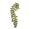
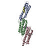
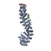
- PDB-1jpp: The Structure of a beta-Catenin Binding Repeat from Adenomatous P... -
+ Open data

- Basic information
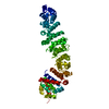
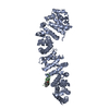
| Entry | Database: PDB / ID: 1jpp | ||||||
|---|---|---|---|---|---|---|---|
| Title | The Structure of a beta-Catenin Binding Repeat from Adenomatous Polyposis Coli (APC) in Complex with beta-Catenin | ||||||
 Components Components |
| ||||||
 Keywords Keywords | CELL ADHESION / Disease mutation / Anti-oncogene | ||||||
| Function / homology |  Function and homology information Function and homology informationlung cell differentiation / epicardium-derived cardiac vascular smooth muscle cell differentiation / mesenchyme morphogenesis / RUNX3 regulates WNT signaling / Regulation of CDH11 function / cardiac vascular smooth muscle cell differentiation / Regulation of MITF-M-dependent genes involved in cell cycle and proliferation / Beta-catenin phosphorylation cascade / APC truncation mutants are not K63 polyubiquitinated / Apoptotic cleavage of cell adhesion proteins ...lung cell differentiation / epicardium-derived cardiac vascular smooth muscle cell differentiation / mesenchyme morphogenesis / RUNX3 regulates WNT signaling / Regulation of CDH11 function / cardiac vascular smooth muscle cell differentiation / Regulation of MITF-M-dependent genes involved in cell cycle and proliferation / Beta-catenin phosphorylation cascade / APC truncation mutants are not K63 polyubiquitinated / Apoptotic cleavage of cell adhesion proteins / hair cycle process / Disassembly of the destruction complex and recruitment of AXIN to the membrane / endoderm formation / LRR FLII-interacting protein 1 (LRRFIP1) activates type I IFN production / positive regulation of epithelial cell differentiation / mesenchyme development / Formation of the beta-catenin:TCF transactivating complex / trachea morphogenesis / Regulation of CDH1 posttranslational processing and trafficking to plasma membrane / metanephros morphogenesis / positive regulation of heparan sulfate proteoglycan biosynthetic process / lung induction / positive regulation of branching involved in lung morphogenesis / cranial ganglion development / renal vesicle formation / renal inner medulla development / renal outer medulla development / nephron tubule formation / beta-catenin-ICAT complex / regulation of epithelial cell differentiation / genitalia morphogenesis / neural plate development / glial cell fate determination / astrocyte-dopaminergic neuron signaling / VEGFR2 mediated vascular permeability / oviduct development / beta-catenin-TCF7L2 complex / regulation of nephron tubule epithelial cell differentiation / regulation of secondary heart field cardioblast proliferation / regulation of timing of anagen / Schwann cell proliferation / negative regulation of mitotic cell cycle, embryonic / central nervous system vasculogenesis / TCF dependent signaling in response to WNT / negative regulation of mesenchymal to epithelial transition involved in metanephros morphogenesis / embryonic skeletal limb joint morphogenesis / regulation of centriole-centriole cohesion / Adherens junctions interactions / Deactivation of the beta-catenin transactivating complex / regulation of centromeric sister chromatid cohesion / animal organ development / negative regulation of cell cycle G1/S phase transition / embryonic axis specification / morphogenesis of embryonic epithelium / Ca2+ pathway / lens morphogenesis in camera-type eye / Scrib-APC-beta-catenin complex / beta-catenin-TCF complex / RHO GTPases activate IQGAPs / negative regulation of cyclin-dependent protein serine/threonine kinase activity / synaptic vesicle clustering / Degradation of beta-catenin by the destruction complex / endodermal cell fate commitment / proximal/distal pattern formation / Regulation of CDH1 Function / Degradation of CDH1 / dorsal/ventral axis specification / dorsal root ganglion development / ventricular compact myocardium morphogenesis / gamma-catenin binding / positive regulation of fibroblast growth factor receptor signaling pathway / regulation of attachment of spindle microtubules to kinetochore / endothelial tube morphogenesis / delta-catenin binding / sympathetic ganglion development / positive regulation of endothelial cell differentiation / layer formation in cerebral cortex / mesenchymal to epithelial transition / ectoderm development / presynaptic active zone cytoplasmic component / hindbrain development / positive regulation of skeletal muscle tissue development / fungiform papilla formation / positive regulation of determination of dorsal identity / regulation of protein localization to cell surface / fascia adherens / embryonic foregut morphogenesis / positive regulation of odontoblast differentiation / smooth muscle cell differentiation / positive regulation of pseudopodium assembly / mesenchymal cell proliferation involved in lung development / alpha-catenin binding / cellular response to indole-3-methanol / regulation of epithelial to mesenchymal transition / regulation of calcium ion import / histone methyltransferase binding / positive regulation of protein localization to centrosome / cell projection membrane / positive regulation of homotypic cell-cell adhesion / negative regulation of oligodendrocyte differentiation Similarity search - Function | ||||||
| Biological species |  | ||||||
| Method |  X-RAY DIFFRACTION / SYNCHROTRON / MOLECULAR REPLACEMENT / Resolution: 3.1 Å X-RAY DIFFRACTION / SYNCHROTRON / MOLECULAR REPLACEMENT / Resolution: 3.1 Å | ||||||
 Authors Authors | Spink, K.E. / Fridman, S.G. / Weis, W.I. | ||||||
 Citation Citation | Journal: EMBO J. / Year: 2001 Title: Molecular mechanisms of beta-catenin recognition by adenomatous polyposis coli revealed by the structure of an APC-beta-catenin complex. Authors: Eklof Spink, K. / Fridman, S.G. / Weis, W.I. #1: Journal: Cell(Cambridge,Mass.) / Year: 2001Title: The structure of the beta-catenin/E-cadherin complex and the molecular basis of diverse ligand recognition by beta-catenin Authors: Huber, A.H. / Weis, W.I. #2: Journal: Cell(Cambridge,Mass.) / Year: 2000Title: CRYSTAL STRUCTURE OF A BETA-CATENIN/TCF COMPLEX Authors: Graham, T.A. / Weaver, C. / Mao, F. / Kimelman, D. / Xu, W. #3: Journal: Cell(Cambridge,Mass.) / Year: 1997Title: THREE-DIMENSIONAL STRUCTURE OF THE ARMADILLO REPEAT REGION OF BETA-CATENIN Authors: Huber, A.H. / Nelson, W.J. / Weis, W.I. | ||||||
| History |
|
- Structure visualization
| Structure viewer | Molecule: MolmilJmol/JSmol |
|---|
- Downloads & links
-Download
| PDBx/mmCIF format | 1jpp.cif.gz | 203.5 KB | Display | PDBx/mmCIF format |
|---|---|---|---|---|
| PDB format | pdb1jpp.ent.gz | 161.1 KB | Display | PDB format |
| PDBx/mmJSON format | 1jpp.json.gz | Tree view | PDBx/mmJSON format | |
| Others |  Other downloads Other downloads |
-Validation report
| Arichive directory | https://data.pdbj.org/pub/pdb/validation_reports/jp/1jppftp://data.pdbj.org/pub/pdb/validation_reports/jp/1jpp | HTTPS FTP |
|---|
-Related structure data
| Related structure data |  1g3jS S: Starting model for refinement |
|---|---|
| Similar structure data |


-Links
 PDBj
PDBj


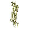
- Assembly
| Deposited unit | 
| ||||||||
|---|---|---|---|---|---|---|---|---|---|
| 1 | 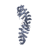
| ||||||||
| 2 | 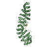
| ||||||||
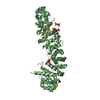
| Unit cell |
|
-Components
| #1: Protein | Mass: 58844.117 Da / Num. of mol.: 2 Source method: isolated from a genetically manipulated source Source: (gene. exp.)  #2: Protein/peptide | Mass: 1786.911 Da / Num. of mol.: 2 / Source method: obtained synthetically Details: This peptide was chemically synthesized. The sequence of the peptide is naturally found in Homo sapiens (human). References: UniProt: P25054 #3: Chemical | ChemComp-GOL / |   Mass: 92.094 Da / Num. of mol.: 1 / Source method: obtained synthetically / Formula: C3H8O3 Mass: 92.094 Da / Num. of mol.: 1 / Source method: obtained synthetically / Formula: C3H8O3#4: Water | ChemComp-HOH / |  Mass: 18.015 Da / Num. of mol.: 72 / Source method: isolated from a natural source / Formula: H2O Mass: 18.015 Da / Num. of mol.: 72 / Source method: isolated from a natural source / Formula: H2O |
|---|
-Experimental details
-Experiment
| Experiment | Method: X-RAY DIFFRACTION / Number of used crystals: 1 |
|---|
- Sample preparation
| Crystal | Density Matthews: 2.58 Å3/Da / Density % sol: 52.23 % | |||||||||||||||||||||||||||||||||||||||||||||||||
|---|---|---|---|---|---|---|---|---|---|---|---|---|---|---|---|---|---|---|---|---|---|---|---|---|---|---|---|---|---|---|---|---|---|---|---|---|---|---|---|---|---|---|---|---|---|---|---|---|---|---|
| Crystal grow | Temperature: 293 K / Method: vapor diffusion, hanging drop / pH: 6.2 Details: PEG 8000, sodium potassium phosphate, sodium chloride, DTT, pH 6.2, VAPOR DIFFUSION, HANGING DROP, temperature 293K | |||||||||||||||||||||||||||||||||||||||||||||||||
| Crystal grow | *PLUS Temperature: 20 ℃ / Method: vapor diffusion, hanging drop | |||||||||||||||||||||||||||||||||||||||||||||||||
| Components of the solutions | *PLUS
|
-Data collection
| Diffraction | Mean temperature: 100 K |
|---|---|
| Diffraction source | Source: SYNCHROTRON / Site: SSRL  / Beamline: BL11-1 / Wavelength: 0.97 Å / Beamline: BL11-1 / Wavelength: 0.97 Å |
| Detector | Type: ADSC QUANTUM 4 / Detector: CCD / Date: Mar 8, 2001 |
| Radiation | Monochromator: curved crystal / Protocol: SINGLE WAVELENGTH / Monochromatic (M) / Laue (L): M / Scattering type: x-ray |
| Radiation wavelength | Wavelength: 0.97 Å / Relative weight: 1 |
| Reflection | Resolution: 3.1→28.3 Å / Num. all: 21958 / Num. obs: 19916 / % possible obs: 90.7 % / Observed criterion σ(I): -3 / Redundancy: 1.47 % / Biso Wilson estimate: 51 Å2 / Rmerge(I) obs: 0.082 |
| Reflection shell | Resolution: 3.1→3.21 Å / Redundancy: 1.39 % / Rmerge(I) obs: 0.242 / Num. unique all: 1937 / % possible all: 87.8 |
| Reflection | *PLUS Lowest resolution: 30 Å |
| Reflection shell | *PLUS % possible obs: 87.8 % |
- Processing
| Software |
| ||||||||||||||||||||||||||||||||||||||||
|---|---|---|---|---|---|---|---|---|---|---|---|---|---|---|---|---|---|---|---|---|---|---|---|---|---|---|---|---|---|---|---|---|---|---|---|---|---|---|---|---|---|
| Refinement | Method to determine structure: MOLECULAR REPLACEMENT Starting model: pdb entry 1G3J Resolution: 3.1→28.27 Å / Rfactor Rfree error: 0.009 / Data cutoff high absF: 1211274.61 / Data cutoff low absF: 0 / Isotropic thermal model: RESTRAINED / Cross valid method: THROUGHOUT / σ(F): 0 / Stereochemistry target values: Engh & Huber
| ||||||||||||||||||||||||||||||||||||||||
| Solvent computation | Solvent model: FLAT MODEL / Bsol: 50.503 Å2 / ksol: 0.320417 e/Å3 | ||||||||||||||||||||||||||||||||||||||||
| Displacement parameters | Biso mean: 28.9 Å2
| ||||||||||||||||||||||||||||||||||||||||
| Refine analyze |
| ||||||||||||||||||||||||||||||||||||||||
| Refinement step | Cycle: LAST / Resolution: 3.1→28.27 Å
| ||||||||||||||||||||||||||||||||||||||||
| Refine LS restraints |
| ||||||||||||||||||||||||||||||||||||||||
| LS refinement shell | Resolution: 3.1→3.21 Å / Rfactor Rfree error: 0.038 / Total num. of bins used: 10
| ||||||||||||||||||||||||||||||||||||||||
| Xplor file |
| ||||||||||||||||||||||||||||||||||||||||
| Software | *PLUS Name: CNS / Version: 1 / Classification: refinement | ||||||||||||||||||||||||||||||||||||||||
| Refinement | *PLUS σ(F): 0 / % reflection Rfree: 5.1 % | ||||||||||||||||||||||||||||||||||||||||
| Solvent computation | *PLUS | ||||||||||||||||||||||||||||||||||||||||
| Displacement parameters | *PLUS Biso mean: 28.9 Å2 | ||||||||||||||||||||||||||||||||||||||||
| Refine LS restraints | *PLUS
| ||||||||||||||||||||||||||||||||||||||||
| LS refinement shell | *PLUS Rfactor Rfree: 0.394 / % reflection Rfree: 6.2 % / Rfactor Rwork: 0.347 |
