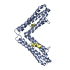
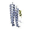
Entry Database : PDB / ID : 3b71Title CD4 endocytosis motif bound to the Focal Adhesion Targeting (FAT) domain of the Focal Adhesion Kinase Focal adhesion kinase 1 T-cell surface glycoprotein CD4 Keywords / / / / / / / / / / / / / / / / / Function / homology Function Domain/homology Component
/ / / / / / / / / / / / / / / / / / / / / / / / / / / / / / / / / / / / / / / / / / / / / / / / / / / / / / / / / / / / / / / / / / / / / / / / / / / / / / / / / / / / / / / / / / / / / / / / / / / / / / / / / / / / / / / / / / / / / / / / / / / / / / / / / / / / / / / / / / / / / / / / / / / / / / / / / / / / / / / / / / / / / / / / / / / Biological species Homo sapiens (human)Method / / / Resolution : 2.82 Å Authors Garron, M.-L. / Arold, S.T. Journal : J.Mol.Biol. / Year : 2008Title : Structural basis for the interaction between focal adhesion kinase and CD4.Authors : Garron, M.L. / Arthos, J. / Guichou, J.F. / McNally, J. / Cicala, C. / Arold, S.T. History Deposition Oct 30, 2007 Deposition site / Processing site Revision 1.0 Jan 1, 2008 Provider / Type Revision 1.1 Jul 13, 2011 Group / Version format complianceRevision 1.2 Aug 30, 2023 Group / Database references / Refinement descriptionCategory chem_comp_atom / chem_comp_bond ... chem_comp_atom / chem_comp_bond / database_2 / pdbx_initial_refinement_model Item / _database_2.pdbx_database_accession
Show all Show less
 Movie
Movie Controller

 Yorodumi
 Open data
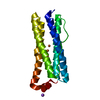
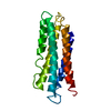
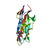
 Basic information
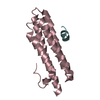
 Components
 Keywords
 Function and homology information
 Homo sapiens (human)
 X-RAY DIFFRACTION /
  Authors
 Citation
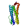
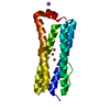
 Structure visualization
 Downloads & links
 Other downloads


 PDBj
PDBj


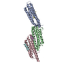
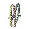
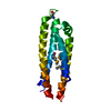
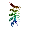
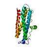
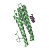
 Assembly
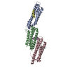

 Sample preparation
 / Beamline: BM30A / Wavelength: 0.97982 Å
 Processing