+ Open data
- Basic information
| Entry | Database: PDB / ID: 7shf | |||||||||
|---|---|---|---|---|---|---|---|---|---|---|
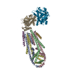
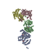
| Title | Cryo-EM structure of GPR158 coupled to the RGS7-Gbeta5 complex | |||||||||
 Components Components |
| |||||||||
 Keywords Keywords | SIGNALING PROTEIN / Receptor / complex | |||||||||
| Function / homology |  Function and homology information Function and homology informationG protein-coupled glycine receptor activity / dendrite terminus / G beta:gamma signalling through PLC beta / Presynaptic function of Kainate receptors / Prostacyclin signalling through prostacyclin receptor / Inactivation, recovery and regulation of the phototransduction cascade / G alpha (z) signalling events / Glucagon-type ligand receptors / G beta:gamma signalling through PI3Kgamma / G beta:gamma signalling through CDC42 ...G protein-coupled glycine receptor activity / dendrite terminus / G beta:gamma signalling through PLC beta / Presynaptic function of Kainate receptors / Prostacyclin signalling through prostacyclin receptor / Inactivation, recovery and regulation of the phototransduction cascade / G alpha (z) signalling events / Glucagon-type ligand receptors / G beta:gamma signalling through PI3Kgamma / G beta:gamma signalling through CDC42 / dark adaptation / light adaption / G-protein gamma-subunit binding / Adrenaline,noradrenaline inhibits insulin secretion / ADP signalling through P2Y purinoceptor 12 / Cooperation of PDCL (PhLP1) and TRiC/CCT in G-protein beta folding / G beta:gamma signalling through BTK / Thromboxane signalling through TP receptor / Thrombin signalling through proteinase activated receptors (PARs) / Activation of G protein gated Potassium channels / Inhibition of voltage gated Ca2+ channels via Gbeta/gamma subunits / negative regulation of voltage-gated calcium channel activity / G alpha (s) signalling events / G-protein activation / Ca2+ pathway / G alpha (12/13) signalling events / Extra-nuclear estrogen signaling / G alpha (q) signalling events / Vasopressin regulates renal water homeostasis via Aquaporins / GPER1 signaling / Glucagon-like Peptide-1 (GLP1) regulates insulin secretion / rod photoreceptor outer segment / cell tip / High laminar flow shear stress activates signaling by PIEZO1 and PECAM1:CDH5:KDR in endothelial cells / ADP signalling through P2Y purinoceptor 1 / G alpha (i) signalling events / regulation of G protein-coupled receptor signaling pathway / negative regulation of G protein-coupled receptor signaling pathway / G protein-coupled dopamine receptor signaling pathway / positive regulation of neurotransmitter secretion / regulation of synapse organization / parallel fiber to Purkinje cell synapse / regulation of postsynaptic membrane potential / G-protein alpha-subunit binding / positive regulation of GTPase activity / photoreceptor inner segment / GTPase activator activity / protein localization to plasma membrane / cell projection / enzyme activator activity / postsynaptic density membrane / brain development / cognition / transmembrane signaling receptor activity / Cooperation of PDCL (PhLP1) and TRiC/CCT in G-protein beta folding / myelin sheath / G-protein beta-subunit binding / presynapse / protein-folding chaperone binding / presynaptic membrane / G alpha (i) signalling events / postsynaptic membrane / neuron projection / intracellular signal transduction / G protein-coupled receptor signaling pathway / GTPase activity / synapse / dendrite / glutamatergic synapse / nucleus / plasma membrane / cytosol / cytoplasm Similarity search - Function | |||||||||
| Biological species |  Homo sapiens (human) Homo sapiens (human) | |||||||||
| Method | ELECTRON MICROSCOPY / single particle reconstruction / cryo EM / Resolution: 3.4 Å | |||||||||
 Authors Authors | Patil, D.N. / Singh, S. / Singh, A.K. / Martemyanov, K.A. | |||||||||
| Funding support |  United States, 1items United States, 1items
| |||||||||
 Citation Citation |  Journal: Science / Year: 2022 Journal: Science / Year: 2022Title: Cryo-EM structure of human GPR158 receptor coupled to the RGS7-Gβ5 signaling complex. Authors: Dipak N Patil / Shikha Singh / Thibaut Laboute / Timothy S Strutzenberg / Xingyu Qiu / Di Wu / Scott J Novick / Carol V Robinson / Patrick R Griffin / John F Hunt / Tina Izard / Appu K Singh ...Authors: Dipak N Patil / Shikha Singh / Thibaut Laboute / Timothy S Strutzenberg / Xingyu Qiu / Di Wu / Scott J Novick / Carol V Robinson / Patrick R Griffin / John F Hunt / Tina Izard / Appu K Singh / Kirill A Martemyanov /    Abstract: GPR158 is an orphan G protein–coupled receptor (GPCR) highly expressed in the brain, where it controls synapse formation and function. GPR158 has also been implicated in depression, carcinogenesis, ...GPR158 is an orphan G protein–coupled receptor (GPCR) highly expressed in the brain, where it controls synapse formation and function. GPR158 has also been implicated in depression, carcinogenesis, and cognition. However, the structural organization and signaling mechanisms of GPR158 are largely unknown. We used single-particle cryo–electron microscopy (cryo-EM) to determine the structures of human GPR158 alone and bound to an RGS signaling complex. The structures reveal a homodimeric organization stabilized by a pair of phospholipids and the presence of an extracellular Cache domain, an unusual ligand-binding domain in GPCRs. We further demonstrate the structural basis of GPR158 coupling to RGS7-Gβ5. Together, these results provide insights into the unusual biology of orphan receptors and the formation of GPCR-RGS complexes. | |||||||||
| History |
|
- Structure visualization
| Movie |
 Movie viewer Movie viewer |
|---|---|
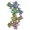
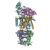
| Structure viewer | Molecule:  Molmil Molmil Jmol/JSmol Jmol/JSmol |
- Downloads & links
- Download
| PDBx/mmCIF format |  7shf.cif.gz 7shf.cif.gz | 322 KB | Display |  PDBx/mmCIF format PDBx/mmCIF format |
|---|---|---|---|---|
| PDB format |  pdb7shf.ent.gz pdb7shf.ent.gz | 258.9 KB | Display |  PDB format PDB format |
| PDBx/mmJSON format |  7shf.json.gz 7shf.json.gz | Tree view |  PDBx/mmJSON format PDBx/mmJSON format | |
| Others |  Other downloads Other downloads |
-Validation report
| Summary document |  7shf_validation.pdf.gz 7shf_validation.pdf.gz | 1.7 MB | Display |  wwPDB validaton report wwPDB validaton report |
|---|---|---|---|---|
| Full document |  7shf_full_validation.pdf.gz 7shf_full_validation.pdf.gz | 1.7 MB | Display | |
| Data in XML |  7shf_validation.xml.gz 7shf_validation.xml.gz | 48.9 KB | Display | |
| Data in CIF |  7shf_validation.cif.gz 7shf_validation.cif.gz | 70.8 KB | Display | |
| Arichive directory |  https://data.pdbj.org/pub/pdb/validation_reports/sh/7shf https://data.pdbj.org/pub/pdb/validation_reports/sh/7shf ftp://data.pdbj.org/pub/pdb/validation_reports/sh/7shf ftp://data.pdbj.org/pub/pdb/validation_reports/sh/7shf | HTTPS FTP |
-Related structure data
| Related structure data |  25126MC  7sheC M: map data used to model this data C: citing same article ( |
|---|---|
| Similar structure data |
- Links
Links
- Assembly
| Deposited unit | 
|
|---|---|
| 1 |
|
- Components
-Protein , 3 types, 4 molecules CDBA
| #1: Protein | Mass: 54761.859 Da / Num. of mol.: 1 Source method: isolated from a genetically manipulated source Source: (gene. exp.)  Homo sapiens (human) / Gene: RGS7 / Production host: Homo sapiens (human) / Gene: RGS7 / Production host:  Homo sapiens (human) / References: UniProt: P49802 Homo sapiens (human) / References: UniProt: P49802 |
|---|---|
| #2: Protein | Mass: 38778.602 Da / Num. of mol.: 1 Source method: isolated from a genetically manipulated source Source: (gene. exp.)   Homo sapiens (human) / References: UniProt: P62881 Homo sapiens (human) / References: UniProt: P62881 |
| #3: Protein | Mass: 88251.633 Da / Num. of mol.: 2 Source method: isolated from a genetically manipulated source Source: (gene. exp.)  Homo sapiens (human) / Gene: GPR158, KIAA1136 / Production host: Homo sapiens (human) / Gene: GPR158, KIAA1136 / Production host:  Homo sapiens (human) / References: UniProt: Q5T848 Homo sapiens (human) / References: UniProt: Q5T848 |
-Non-polymers , 3 types, 24 molecules 


| #4: Chemical | ChemComp-CLR / #5: Chemical | ChemComp-EIJ / ( | #6: Chemical | ChemComp-PEE / | |
|---|
-Details
| Has ligand of interest | N |
|---|
-Experimental details
-Experiment
| Experiment | Method: ELECTRON MICROSCOPY |
|---|---|
| EM experiment | Aggregation state: PARTICLE / 3D reconstruction method: single particle reconstruction |
- Sample preparation
| Component | Name: GPCR complex / Type: COMPLEX / Entity ID: #1-#3 / Source: MULTIPLE SOURCES |
|---|---|
| Molecular weight | Value: 0.270 MDa / Experimental value: NO |
| Source (natural) | Organism:  Homo sapiens (human) Homo sapiens (human) |
| Source (recombinant) | Organism:  Homo sapiens (human) Homo sapiens (human) |
| Buffer solution | pH: 8 |
| Specimen | Conc.: 0.3 mg/ml / Embedding applied: NO / Shadowing applied: NO / Staining applied: NO / Vitrification applied: YES |
| Vitrification | Cryogen name: ETHANE / Humidity: 100 % / Chamber temperature: 277 K |
- Electron microscopy imaging
| Experimental equipment |  Model: Titan Krios / Image courtesy: FEI Company |
|---|---|
| Microscopy | Model: FEI TITAN KRIOS |
| Electron gun | Electron source:  FIELD EMISSION GUN / Accelerating voltage: 300 kV / Illumination mode: OTHER FIELD EMISSION GUN / Accelerating voltage: 300 kV / Illumination mode: OTHER |
| Electron lens | Mode: OTHER / Nominal defocus max: 2500 nm / Nominal defocus min: 1500 nm |
| Image recording | Electron dose: 40 e/Å2 / Film or detector model: GATAN K3 (6k x 4k) |
- Processing
| EM software | Name: cryoSPARC / Category: 3D reconstruction |
|---|---|
| CTF correction | Type: PHASE FLIPPING AND AMPLITUDE CORRECTION |
| Symmetry | Point symmetry: C1 (asymmetric) |
| 3D reconstruction | Resolution: 3.4 Å / Resolution method: FSC 0.143 CUT-OFF / Num. of particles: 151954 / Symmetry type: POINT |
 Movie
Movie Controller





 PDBj
PDBj


