Movie
Movie Controller


+ Open data

- Basic information
| Entry | Database: EMDB / ID: EMD-25126 | |||||||||
|---|---|---|---|---|---|---|---|---|---|---|
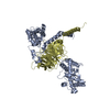
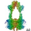
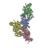
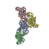
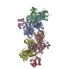
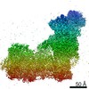
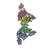
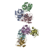
| Title | Cryo-EM structure of GPR158 coupled to the RGS7-Gbeta5 complex | |||||||||
 Map data Map data | ||||||||||
 Sample Sample |
| |||||||||
| Function / homology |  Function and homology information Function and homology informationG protein-coupled glycine receptor activity / dendrite terminus / Inactivation, recovery and regulation of the phototransduction cascade / G beta:gamma signalling through PLC beta / Presynaptic function of Kainate receptors / Prostacyclin signalling through prostacyclin receptor / G alpha (z) signalling events / Glucagon-type ligand receptors / G beta:gamma signalling through PI3Kgamma / G beta:gamma signalling through CDC42 ...G protein-coupled glycine receptor activity / dendrite terminus / Inactivation, recovery and regulation of the phototransduction cascade / G beta:gamma signalling through PLC beta / Presynaptic function of Kainate receptors / Prostacyclin signalling through prostacyclin receptor / G alpha (z) signalling events / Glucagon-type ligand receptors / G beta:gamma signalling through PI3Kgamma / G beta:gamma signalling through CDC42 / Adrenaline,noradrenaline inhibits insulin secretion / ADP signalling through P2Y purinoceptor 12 / Cooperation of PDCL (PhLP1) and TRiC/CCT in G-protein beta folding / Thromboxane signalling through TP receptor / G beta:gamma signalling through BTK / Thrombin signalling through proteinase activated receptors (PARs) / Activation of G protein gated Potassium channels / Inhibition of voltage gated Ca2+ channels via Gbeta/gamma subunits / light adaption / dark adaptation / G-protein gamma-subunit binding / G alpha (s) signalling events / Ca2+ pathway / G-protein activation / Extra-nuclear estrogen signaling / G alpha (12/13) signalling events / G alpha (q) signalling events / Vasopressin regulates renal water homeostasis via Aquaporins / GPER1 signaling / Glucagon-like Peptide-1 (GLP1) regulates insulin secretion / ADP signalling through P2Y purinoceptor 1 / High laminar flow shear stress activates signaling by PIEZO1 and PECAM1:CDH5:KDR in endothelial cells / cell tip / G alpha (i) signalling events / regulation of G protein-coupled receptor signaling pathway / GTPase activating protein binding / G protein-coupled dopamine receptor signaling pathway / negative regulation of G protein-coupled receptor signaling pathway / positive regulation of neurotransmitter secretion / regulation of synapse organization / parallel fiber to Purkinje cell synapse / regulation of postsynaptic membrane potential / positive regulation of GTPase activity / photoreceptor outer segment / G-protein alpha-subunit binding / photoreceptor inner segment / GTPase activator activity / protein localization to plasma membrane / brain development / postsynaptic density membrane / cognition / enzyme activator activity / transmembrane signaling receptor activity / myelin sheath / Cooperation of PDCL (PhLP1) and TRiC/CCT in G-protein beta folding / heterotrimeric G-protein complex / G-protein beta-subunit binding / protein-folding chaperone binding / presynapse / signaling receptor complex adaptor activity / cell body / presynaptic membrane / G alpha (i) signalling events / postsynaptic membrane / neuron projection / intracellular signal transduction / G protein-coupled receptor signaling pathway / GTPase activity / synapse / dendrite / glutamatergic synapse / protein-containing complex / nucleus / plasma membrane / cytoplasm / cytosol Similarity search - Function | |||||||||
| Biological species |  Homo sapiens (human) / Homo sapiens (human) /  | |||||||||
| Method | single particle reconstruction / cryo EM / Resolution: 3.4 Å | |||||||||
 Authors Authors | Patil DN / Singh S / Singh AK / Martemyanov KA | |||||||||
| Funding support |  United States, 1 items United States, 1 items
| |||||||||
 Citation Citation | Journal: Science / Year: 2022 Title: Cryo-EM structure of human GPR158 receptor coupled to the RGS7-Gβ5 signaling complex. Authors: Dipak N Patil / Shikha Singh / Thibaut Laboute / Timothy S Strutzenberg / Xingyu Qiu / Di Wu / Scott J Novick / Carol V Robinson / Patrick R Griffin / John F Hunt / Tina Izard / Appu K Singh ...Authors: Dipak N Patil / Shikha Singh / Thibaut Laboute / Timothy S Strutzenberg / Xingyu Qiu / Di Wu / Scott J Novick / Carol V Robinson / Patrick R Griffin / John F Hunt / Tina Izard / Appu K Singh / Kirill A Martemyanov /   Abstract: GPR158 is an orphan G protein–coupled receptor (GPCR) highly expressed in the brain, where it controls synapse formation and function. GPR158 has also been implicated in depression, carcinogenesis, ...GPR158 is an orphan G protein–coupled receptor (GPCR) highly expressed in the brain, where it controls synapse formation and function. GPR158 has also been implicated in depression, carcinogenesis, and cognition. However, the structural organization and signaling mechanisms of GPR158 are largely unknown. We used single-particle cryo–electron microscopy (cryo-EM) to determine the structures of human GPR158 alone and bound to an RGS signaling complex. The structures reveal a homodimeric organization stabilized by a pair of phospholipids and the presence of an extracellular Cache domain, an unusual ligand-binding domain in GPCRs. We further demonstrate the structural basis of GPR158 coupling to RGS7-Gβ5. Together, these results provide insights into the unusual biology of orphan receptors and the formation of GPCR-RGS complexes. | |||||||||
| History |
|
- Structure visualization
| Movie |
Movie viewer |
|---|---|
| Structure viewer | EM map: SurfViewMolmilJmol/JSmol |
| Supplemental images |

- Downloads & links
-EMDB archive
| Map data | emd_25126.map.gz | 230.2 MB | EMDB map data format | |
|---|---|---|---|---|
| Header (meta data) | emd-25126-v30.xmlemd-25126.xml | 15.3 KB 15.3 KB | Display Display | EMDB header |
| Images | 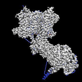 emd_25126.png emd_25126.png | 63 KB | ||
| Archive directory |  http://ftp.pdbj.org/pub/emdb/structures/EMD-25126ftp://ftp.pdbj.org/pub/emdb/structures/EMD-25126 http://ftp.pdbj.org/pub/emdb/structures/EMD-25126ftp://ftp.pdbj.org/pub/emdb/structures/EMD-25126 | HTTPS FTP |
-Related structure data
| Related structure data | 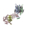 7shfMC 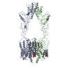 7sheC C: citing same article ( M: atomic model generated by this map |
|---|---|
| Similar structure data |
-Links
| EMDB pages | EMDB (EBI/PDBe) / EMDataResource |
|---|---|
| Related items in Molecule of the Month |


-Map
| File | Download / File: emd_25126.map.gz / Format: CCP4 / Size: 244.1 MB / Type: IMAGE STORED AS FLOATING POINT NUMBER (4 BYTES) | ||||||||||||||||||||||||||||||||||||||||||||||||||||||||||||||||||||
|---|---|---|---|---|---|---|---|---|---|---|---|---|---|---|---|---|---|---|---|---|---|---|---|---|---|---|---|---|---|---|---|---|---|---|---|---|---|---|---|---|---|---|---|---|---|---|---|---|---|---|---|---|---|---|---|---|---|---|---|---|---|---|---|---|---|---|---|---|---|
| Projections & slices | Image control
Images are generated by Spider. | ||||||||||||||||||||||||||||||||||||||||||||||||||||||||||||||||||||
| Voxel size | X=Y=Z: 0.873 Å | ||||||||||||||||||||||||||||||||||||||||||||||||||||||||||||||||||||
| Density |
| ||||||||||||||||||||||||||||||||||||||||||||||||||||||||||||||||||||
| Symmetry | Space group: 1 | ||||||||||||||||||||||||||||||||||||||||||||||||||||||||||||||||||||
| Details | EMDB XML:
CCP4 map header:
| ||||||||||||||||||||||||||||||||||||||||||||||||||||||||||||||||||||
 Z (Sec.)
 Y (Row.)
 X (Col.)


-Supplemental data
- Sample components
-Entire : GPCR complex
| Entire | Name: GPCR complex |
|---|---|
| Components |
|
-Supramolecule #1: GPCR complex
| Supramolecule | Name: GPCR complex / type: complex / ID: 1 / Parent: 0 / Macromolecule list: #1-#3 |
|---|---|
| Source (natural) | Organism: Homo sapiens (human) |
| Molecular weight | Theoretical: 270 KDa |
-Macromolecule #1: Isoform 2 of Regulator of G-protein signaling 7
| Macromolecule | Name: Isoform 2 of Regulator of G-protein signaling 7 / type: protein_or_peptide / ID: 1 / Number of copies: 1 / Enantiomer: LEVO |
|---|---|
| Source (natural) | Organism: Homo sapiens (human) |
| Molecular weight | Theoretical: 54.761859 KDa |
| Recombinant expression | Organism: Homo sapiens (human) |
| Sequence | String: MAQGNNYGQT SNGVADESPN MLVYRKMEDV IARMQDEKNG IPIRTVKSFL SKIPSVFSGS DIVQWLIKNL TIEDPVEALH LGTLMAAHG YFFPISDHVL TLKDDGTFYR FQTPYFWPSN CWEPENTDYA VYLCKRTMQN KARLELADYE AESLARLQRA F ARKWEFIF ...String: MAQGNNYGQT SNGVADESPN MLVYRKMEDV IARMQDEKNG IPIRTVKSFL SKIPSVFSGS DIVQWLIKNL TIEDPVEALH LGTLMAAHG YFFPISDHVL TLKDDGTFYR FQTPYFWPSN CWEPENTDYA VYLCKRTMQN KARLELADYE AESLARLQRA F ARKWEFIF MQAEAQAKVD KKRDKIERKI LDSQERAFWD VHRPVPGCVN TTEVDIKKSS RMRNPHKTRK SVYGLQNDIR SH SPTHTPT PETKPPTEDE LQQQIKYWQI QLDRHRLKMS KVADSLLSYT EQYLEYDPFL LPPDPSNPWL SDDTTFWELE ASK EPSQQR VKRWGFGMDE ALKDPVGREQ FLKFLESEFS SENLRFWLAV EDLKKRPIKE VPSRVQEIWQ EFLAPGAPSA INLD SKSYD KTTQNVKEPG RYTFEDAQEH IYKLMKSDSY PRFIRSSAYQ ELLQAKKKGK SLTSKRLTSL AQSY |
-Macromolecule #2: Guanine nucleotide-binding protein subunit beta-5
| Macromolecule | Name: Guanine nucleotide-binding protein subunit beta-5 / type: protein_or_peptide / ID: 2 / Number of copies: 1 / Enantiomer: LEVO |
|---|---|
| Source (natural) | Organism: |
| Molecular weight | Theoretical: 38.778602 KDa |
| Recombinant expression | Organism: Homo sapiens (human) |
| Sequence | String: MATDGLHENE TLASLKSEAE SLKGKLEEER AKLHDVELHQ VAERVEALGQ FVMKTRRTLK GHGNKVLCMD WCKDKRRIVS SSQDGKVIV WDSFTTNKEH AVTMPCTWVM ACAYAPSGCA IACGGLDNKC SVYPLTFDKN ENMAAKKKSV AMHTNYLSAC S FTNSDMQI ...String: MATDGLHENE TLASLKSEAE SLKGKLEEER AKLHDVELHQ VAERVEALGQ FVMKTRRTLK GHGNKVLCMD WCKDKRRIVS SSQDGKVIV WDSFTTNKEH AVTMPCTWVM ACAYAPSGCA IACGGLDNKC SVYPLTFDKN ENMAAKKKSV AMHTNYLSAC S FTNSDMQI LTASGDGTCA LWDVESGQLL QSFHGHGADV LCLDLAPSET GNTFVSGGCD KKAMVWDMRS GQCVQAFETH ES DVNSVRY YPSGDAFASG SDDATCRLYD LRADREVAIY SKESIIFGAS SVDFSLSGRL LFAGYNDYTI NVWDVLKGSR VSI LFGHEN RVSTLRVSPD GTAFCSGSWD HTLRVWA |
-Macromolecule #3: G-protein coupled receptor 158
| Macromolecule | Name: G-protein coupled receptor 158 / type: protein_or_peptide / ID: 3 / Number of copies: 2 / Enantiomer: LEVO |
|---|---|
| Source (natural) | Organism: Homo sapiens (human) |
| Molecular weight | Theoretical: 88.251633 KDa |
| Recombinant expression | Organism: Homo sapiens (human) |
| Sequence | String: MGAMAYPLLL CLLLAQLGLG AVGASRDPQG RPDSPRERTP KGKPHAQQPG RASASDSSAP WSRSTDGTIL AQKLAEEVPM DVASYLYTG DSHQLKRANC SGRYELAGLP GKWPALASAH PSLHRALDTL THATNFLNVM LQSNKSREQN LQDDLDWYQA L VWSLLEGE ...String: MGAMAYPLLL CLLLAQLGLG AVGASRDPQG RPDSPRERTP KGKPHAQQPG RASASDSSAP WSRSTDGTIL AQKLAEEVPM DVASYLYTG DSHQLKRANC SGRYELAGLP GKWPALASAH PSLHRALDTL THATNFLNVM LQSNKSREQN LQDDLDWYQA L VWSLLEGE PSISRAAITF STDSLSAPAP QVFLQATREE SRILLQDLSS SAPHLANATL ETEWFHGLRR KWRPHLHRRG PN QGPRGLG HSWRRKDGLG GDKSHFKWSP PYLECENGSY KPGWLVTLSS AIYGLQPNLV PEFRGVMKVD INLQKVDIDQ CSS DGWFSG THKCHLNNSE CMPIKGLGFV LGAYECICKA GFYHPGVLPV NNFRRRGPDQ HISGSTKDVS EEAYVCLPCR EGCP FCADD SPCFVQEDKY LRLAIISFQA LCMLLDFVSM LVVYHFRKAK SIRASGLILL ETILFGSLLL YFPVVILYFE PSTFR CILL RWARLLGFAT VYGTVTLKLH RVLKVFLSRT AQRIPYMTGG RVMRMLAVIL LVVFWFLIGW TSSVCQNLEK QISLIG QGK TSDHLIFNMC LIDRWDYMTA VAEFLFLLWG VYLCYAVRTV PSAFHEPRYM AVAVHNELII SAIFHTIRFV LASRLQS DW MLMLYFAHTH LTVTVTIGLL LIPKFSHSSN NPRDDIATEA YEDELDMGRS GSYLNSSINS AWSEHSLDPE DIRDELKK L YAQLEIYKRK KMITNNPHLQ KKRCSKKGLG RSIMRRITEI PETVSRQCSK EDKELEVLFQ |
-Macromolecule #4: CHOLESTEROL
| Macromolecule | Name: CHOLESTEROL / type: ligand / ID: 4 / Number of copies: 22 / Formula: CLR |
|---|---|
| Molecular weight | Theoretical: 386.654 Da |
| Chemical component information |  ChemComp-CLR: |
-Macromolecule #5: (2S)-1-{[(S)-hydroxy{[(1s,2R,3R,4R,5S,6S)-2,3,4,5,6-pentahydroxyc...
| Macromolecule | Name: (2S)-1-{[(S)-hydroxy{[(1s,2R,3R,4R,5S,6S)-2,3,4,5,6-pentahydroxycyclohexyl]oxy}phosphoryl]oxy}-3-(octadecanoyloxy)propan-2-yl (5E,8E,11E,14E)-icosa-5,8,11,14-tetraenoate type: ligand / ID: 5 / Number of copies: 1 / Formula: EIJ |
|---|---|
| Molecular weight | Theoretical: 887.128 Da |
| Chemical component information |  ChemComp-EIJ: |
-Macromolecule #6: 1,2-dioleoyl-sn-glycero-3-phosphoethanolamine
| Macromolecule | Name: 1,2-dioleoyl-sn-glycero-3-phosphoethanolamine / type: ligand / ID: 6 / Number of copies: 1 / Formula: PEE |
|---|---|
| Molecular weight | Theoretical: 744.034 Da |
| Chemical component information |  ChemComp-PEE: |
-Experimental details
-Structure determination
| Method | cryo EM |
|---|---|
 Processing Processing | single particle reconstruction |
| Aggregation state | particle |
-Sample preparation
| Concentration | 0.3 mg/mL |
|---|---|
| Buffer | pH: 8 |
| Vitrification | Cryogen name: ETHANE / Chamber humidity: 100 % / Chamber temperature: 277 K |
- Electron microscopy
| Microscope | FEI TITAN KRIOS |
|---|---|
| Image recording | Film or detector model: GATAN K3 (6k x 4k) / Average electron dose: 40.0 e/Å2 |
| Electron beam | Acceleration voltage: 300 kV / Electron source:  FIELD EMISSION GUN FIELD EMISSION GUN |
| Electron optics | Illumination mode: OTHER / Imaging mode: OTHER / Nominal defocus max: 2.5 µm / Nominal defocus min: 1.5 µm |
| Experimental equipment |  Model: Titan Krios / Image courtesy: FEI Company |
-Image processing
| Startup model | Type of model: PDB ENTRY PDB model - PDB ID: Details: GPR158 model taken from ab-initio built for GPR158 apo and RGS7-Gb5 from PDB ID: 6N9G |
|---|---|
| Final reconstruction | Applied symmetry - Point group: C1 (asymmetric) / Resolution.type: BY AUTHOR / Resolution: 3.4 Å / Resolution method: FSC 0.143 CUT-OFF / Software - Name: cryoSPARC / Number images used: 151954 |
| Initial angle assignment | Type: MAXIMUM LIKELIHOOD |
| Final angle assignment | Type: MAXIMUM LIKELIHOOD |