 Movie
Movie Controller

[English] 日本語
 Yorodumi
- PDB-4j56: Structure of Plasmodium falciparum thioredoxin reductase-thioredo... -
+ Open data

- Basic information
| Entry | Database: PDB / ID: 4j56 | ||||||
|---|---|---|---|---|---|---|---|
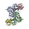
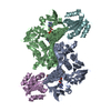
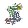
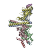
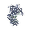
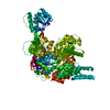
| Title | Structure of Plasmodium falciparum thioredoxin reductase-thioredoxin complex | ||||||
 Components Components |
| ||||||
 Keywords Keywords | OXIDOREDUCTASE / Protein-Protein Complex / Thioredoxin fold / Disulfide reductase / FAD binding / NADPH binding / Thioredoxin binding | ||||||
| Function / homology |  Function and homology information Function and homology informationMetabolism of ingested MeSeO2H into MeSeH / : / TP53 Regulates Metabolic Genes / Detoxification of Reactive Oxygen Species / Interconversion of nucleotide di- and triphosphates / thioredoxin-disulfide reductase (NADPH) / thioredoxin-disulfide reductase (NADPH) activity / protein-disulfide reductase activity / cell redox homeostasis / flavin adenine dinucleotide binding ...Metabolism of ingested MeSeO2H into MeSeH / : / TP53 Regulates Metabolic Genes / Detoxification of Reactive Oxygen Species / Interconversion of nucleotide di- and triphosphates / thioredoxin-disulfide reductase (NADPH) / thioredoxin-disulfide reductase (NADPH) activity / protein-disulfide reductase activity / cell redox homeostasis / flavin adenine dinucleotide binding / mitochondrion / cytoplasm / cytosol Similarity search - Function | ||||||
| Biological species |  | ||||||
| Method |  X-RAY DIFFRACTION / SYNCHROTRON / MOLECULAR REPLACEMENT / Resolution: 2.371 Å X-RAY DIFFRACTION / SYNCHROTRON / MOLECULAR REPLACEMENT / Resolution: 2.371 Å | ||||||
 Authors Authors | Fritz-Wolf, K. / Jortzik, E. / Stumpf, M. / Preuss, J. / Iozef, R. / Rahlfs, S. / Becker, K. | ||||||
 Citation Citation | Journal: J.Mol.Biol. / Year: 2013 Title: Crystal Structure of the Plasmodium falciparum Thioredoxin Reductase-Thioredoxin Complex. Authors: Fritz-Wolf, K. / Jortzik, E. / Stumpf, M. / Preuss, J. / Iozef, R. / Rahlfs, S. / Becker, K. | ||||||
| History |
|
- Structure visualization
| Structure viewer | Molecule: MolmilJmol/JSmol |
|---|
- Downloads & links
-Download
| PDBx/mmCIF format | 4j56.cif.gz | 477.8 KB | Display | PDBx/mmCIF format |
|---|---|---|---|---|
| PDB format | pdb4j56.ent.gz | 391.1 KB | Display | PDB format |
| PDBx/mmJSON format | 4j56.json.gz | Tree view | PDBx/mmJSON format | |
| Others |  Other downloads Other downloads |
-Validation report
| Arichive directory | https://data.pdbj.org/pub/pdb/validation_reports/j5/4j56ftp://data.pdbj.org/pub/pdb/validation_reports/j5/4j56 | HTTPS FTP |
|---|
-Related structure data
| Related structure data |  4j57C  3qfbS S: Starting model for refinement C: citing same article ( |
|---|---|
| Similar structure data |


-Links
 PDBj
PDBj


- Assembly
| Deposited unit | 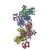
| |||||||||||||||||||||||||||||||||||||||||||||||||||||||||
|---|---|---|---|---|---|---|---|---|---|---|---|---|---|---|---|---|---|---|---|---|---|---|---|---|---|---|---|---|---|---|---|---|---|---|---|---|---|---|---|---|---|---|---|---|---|---|---|---|---|---|---|---|---|---|---|---|---|---|
| 1 | 
| |||||||||||||||||||||||||||||||||||||||||||||||||||||||||
| 2 | 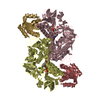
| |||||||||||||||||||||||||||||||||||||||||||||||||||||||||
| Unit cell |
| |||||||||||||||||||||||||||||||||||||||||||||||||||||||||
| Noncrystallographic symmetry (NCS) | NCS domain:
NCS domain segments:
NCS ensembles :
|
-Components
| #1: Protein | Mass: 59752.160 Da / Num. of mol.: 4 / Mutation: C535S Source method: isolated from a genetically manipulated source Source: (gene. exp.) Strain: 3D7 / Gene: trxr2, PFI1170c / Plasmid: pRSETa / Production host:  References: UniProt: P61076, thioredoxin-disulfide reductase (NADPH) #2: Protein | Mass: 12854.468 Da / Num. of mol.: 4 / Mutation: C33S Source method: isolated from a genetically manipulated source Source: (gene. exp.) Strain: 3D7 / Gene: PF14_0545 / Plasmid: pQE30 / Production host: #3: Chemical | ChemComp-FAD /   Mass: 785.550 Da / Num. of mol.: 4 / Source method: obtained synthetically / Formula: C27H33N9O15P2 / Comment: FAD*YM Mass: 785.550 Da / Num. of mol.: 4 / Source method: obtained synthetically / Formula: C27H33N9O15P2 / Comment: FAD*YM#4: Water | ChemComp-HOH / |  Mass: 18.015 Da / Num. of mol.: 215 / Source method: isolated from a natural source / Formula: H2O Mass: 18.015 Da / Num. of mol.: 215 / Source method: isolated from a natural source / Formula: H2OHas protein modification | Y | |
|---|
-Experimental details
-Experiment
| Experiment | Method: X-RAY DIFFRACTION / Number of used crystals: 1 |
|---|
- Sample preparation
| Crystal | Density Matthews: 2.37 Å3/Da / Density % sol: 48 % |
|---|---|
| Crystal grow | Temperature: 295 K / Method: vapor diffusion, hanging drop / pH: 7.6 Details: 20% PEG 8000, pH 7.6, VAPOR DIFFUSION, HANGING DROP, temperature 295K |
-Data collection
| Diffraction | Mean temperature: 100 K |
|---|---|
| Diffraction source | Source: SYNCHROTRON / Site: SLS  / Beamline: X10SA / Wavelength: 0.98696 Å / Beamline: X10SA / Wavelength: 0.98696 Å |
| Detector | Type: MARMOSAIC 225 mm CCD / Detector: CCD / Date: Nov 27, 2008 |
| Radiation | Protocol: SINGLE WAVELENGTH / Monochromatic (M) / Laue (L): M / Scattering type: x-ray |
| Radiation wavelength | Wavelength: 0.98696 Å / Relative weight: 1 |
| Reflection | Resolution: 2.37→50 Å / Num. all: 110173 / Num. obs: 107823 / % possible obs: 97.9 % / Observed criterion σ(F): 0 / Observed criterion σ(I): -3 / Redundancy: 3.7 % / Biso Wilson estimate: 56.97 Å2 / Rsym value: 0.109 / Net I/σ(I): 9.35 |
| Reflection shell | Resolution: 2.37→2.43 Å / Redundancy: 3 % / % possible all: 84.7 |
- Processing
| Software |
| |||||||||||||||||||||||||||||||||||||||||||||||||||||||||||||||||||||||||||||||||||||||||||||||||||||||||||||||||||||||||||||||||||||||||||||||||||||||||||||||||||||||||||||||||||||||||||||||||||||||||||||||||||||||||
|---|---|---|---|---|---|---|---|---|---|---|---|---|---|---|---|---|---|---|---|---|---|---|---|---|---|---|---|---|---|---|---|---|---|---|---|---|---|---|---|---|---|---|---|---|---|---|---|---|---|---|---|---|---|---|---|---|---|---|---|---|---|---|---|---|---|---|---|---|---|---|---|---|---|---|---|---|---|---|---|---|---|---|---|---|---|---|---|---|---|---|---|---|---|---|---|---|---|---|---|---|---|---|---|---|---|---|---|---|---|---|---|---|---|---|---|---|---|---|---|---|---|---|---|---|---|---|---|---|---|---|---|---|---|---|---|---|---|---|---|---|---|---|---|---|---|---|---|---|---|---|---|---|---|---|---|---|---|---|---|---|---|---|---|---|---|---|---|---|---|---|---|---|---|---|---|---|---|---|---|---|---|---|---|---|---|---|---|---|---|---|---|---|---|---|---|---|---|---|---|---|---|---|---|---|---|---|---|---|---|---|---|---|---|---|---|---|---|---|
| Refinement | Method to determine structure: MOLECULAR REPLACEMENT Starting model: 3qfb Resolution: 2.371→19.971 Å / SU ML: 0.37 / σ(F): 1.99 / Phase error: 37.7 / Stereochemistry target values: ML
| |||||||||||||||||||||||||||||||||||||||||||||||||||||||||||||||||||||||||||||||||||||||||||||||||||||||||||||||||||||||||||||||||||||||||||||||||||||||||||||||||||||||||||||||||||||||||||||||||||||||||||||||||||||||||
| Solvent computation | Shrinkage radii: 0.9 Å / VDW probe radii: 1.11 Å / Solvent model: FLAT BULK SOLVENT MODEL | |||||||||||||||||||||||||||||||||||||||||||||||||||||||||||||||||||||||||||||||||||||||||||||||||||||||||||||||||||||||||||||||||||||||||||||||||||||||||||||||||||||||||||||||||||||||||||||||||||||||||||||||||||||||||
| Displacement parameters |
| |||||||||||||||||||||||||||||||||||||||||||||||||||||||||||||||||||||||||||||||||||||||||||||||||||||||||||||||||||||||||||||||||||||||||||||||||||||||||||||||||||||||||||||||||||||||||||||||||||||||||||||||||||||||||
| Refinement step | Cycle: LAST / Resolution: 2.371→19.971 Å
| |||||||||||||||||||||||||||||||||||||||||||||||||||||||||||||||||||||||||||||||||||||||||||||||||||||||||||||||||||||||||||||||||||||||||||||||||||||||||||||||||||||||||||||||||||||||||||||||||||||||||||||||||||||||||
| Refine LS restraints |
| |||||||||||||||||||||||||||||||||||||||||||||||||||||||||||||||||||||||||||||||||||||||||||||||||||||||||||||||||||||||||||||||||||||||||||||||||||||||||||||||||||||||||||||||||||||||||||||||||||||||||||||||||||||||||
| Refine LS restraints NCS |
| |||||||||||||||||||||||||||||||||||||||||||||||||||||||||||||||||||||||||||||||||||||||||||||||||||||||||||||||||||||||||||||||||||||||||||||||||||||||||||||||||||||||||||||||||||||||||||||||||||||||||||||||||||||||||
| LS refinement shell |
|
