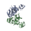
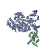
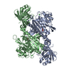
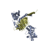
Entry Database : PDB / ID : 6n9gTitle Crystal Structure of RGS7-Gbeta5 dimer Guanine nucleotide-binding protein subunit beta-5 Regulator of G-protein signaling 7 Keywords / / Function / homology Function Domain/homology Component
/ / / / / / / / / / / / / / / / / / / / / / / / / / / / / / / / / / / / / / / / / / / / / / / / / / / / / / / / / / / / / / / / / / / / / / / / / / / / / / / / / / / / / / / / / / / / / / / / / / / / / / / / / / / / / / / / / / / / / / / / / / / Biological species Bos taurus (domestic cattle)Mus musculus (house mouse)Method / / / Resolution : 2.129 Å Authors Patil, D.N. / Rangarajan, E. / Izard, T. / Martemyanov, K.A. Funding support Organization Grant number Country National Institutes of Health/National Institute on Drug Abuse (NIH/NIDA) DA036596
Journal : Elife / Year : 2018Title : Structural organization of a major neuronal G protein regulator, the RGS7-G beta 5-R7BP complex.Authors : Patil, D.N. / Rangarajan, E.S. / Novick, S.J. / Pascal, B.D. / Kojetin, D.J. / Griffin, P.R. / Izard, T. / Martemyanov, K.A. History Deposition Dec 3, 2018 Deposition site / Processing site Revision 1.0 Jan 9, 2019 Provider / Type Revision 1.1 Dec 11, 2019 Group / Category / Item Revision 1.2 Oct 11, 2023 Group / Database references / Refinement descriptionCategory chem_comp_atom / chem_comp_bond ... chem_comp_atom / chem_comp_bond / database_2 / pdbx_initial_refinement_model / software Item / _database_2.pdbx_database_accession / _software.name
Show all Show less
 Movie
Movie Controller


 Open data
 Basic information
 Components
 Keywords
 Function and homology information
 X-RAY DIFFRACTION /
  Authors
 United States, 1items
  Citation
 Structure visualization
 Downloads & links
 Other downloads
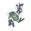


 PDBj
PDBj


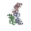
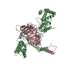
 Assembly
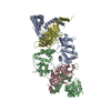

 Spodoptera frugiperda (fall armyworm) / References: UniProt: O46470
 Mass: 18.015 Da / Num. of mol.: 479 / Source method: isolated from a natural source / Formula: H2O
 Sample preparation
 Processing